

Modulating Regional Motor Cortical Excitability with Noninvasive Brain Stimulation Results in Neurochemical Changes in Bilateral Motor Cortices
- PMID: 30030397
- PMCID: PMC6096041
- DOI: 10.1523/JNEUROSCI.2853-17.2018
Modulating Regional Motor Cortical Excitability with Noninvasive Brain Stimulation Results in Neurochemical Changes in Bilateral Motor Cortices
Abstract
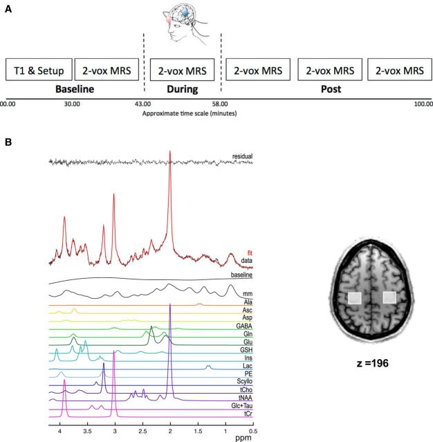
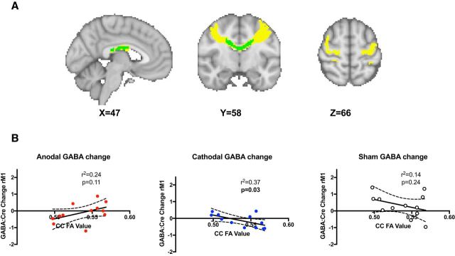
Learning a novel motor skill is dependent both on regional changes within the primary motor cortex (M1) contralateral to the active hand and also on modulation between and within anatomically distant but functionally connected brain regions. Interregional changes are particularly important in functional recovery after stroke, when critical plastic changes underpinning behavioral improvements are observed in both ipsilesional and contralesional M1s. It is increasingly understood that reduction in GABA in the contralateral M1 is necessary to allow learning of a motor task. However, the physiological mechanisms underpinning plasticity within other brain regions, most importantly the ipsilateral M1, are not well understood. Here, we used concurrent two-voxel magnetic resonance spectroscopy to simultaneously quantify changes in neurochemicals within left and right M1s in healthy humans of both sexes in response to transcranial direct current stimulation (tDCS) applied to left M1. We demonstrated a decrease in GABA in both the stimulated (left) and nonstimulated (right) M1 after anodal tDCS, whereas a decrease in GABA was only observed in nonstimulated M1 after cathodal stimulation. This GABA decrease in the nonstimulated M1 during cathodal tDCS was negatively correlated with microstructure of M1:M1 callosal fibers, as quantified by diffusion MRI, suggesting that structural features of these fibers may mediate GABA decrease in the unstimulated region. We found no significant changes in glutamate. Together, these findings shed light on the interactions between the two major network nodes underpinning motor plasticity, offering a potential framework from which to optimize future interventions to improve motor function after stroke.SIGNIFICANCE STATEMENT Learning of new motor skills depends on modulation both within and between brain regions. Here, we use a novel two-voxel magnetic resonance spectroscopy approach to quantify GABA and glutamate changes concurrently within the left and right primary motor cortex (M1) during three commonly used transcranial direct current stimulation montages: anodal, cathodal, and bilateral. We also examined how the neurochemical changes in the unstimulated hemisphere were related to white matter microstructure between the two M1s. Our results provide insights into the neurochemical changes underlying motor plasticity and may therefore assist in the development of further adjunct therapies.
Keywords: DTI; GABA; M1; MRS; plasticity; tDCS.
Copyright © 2018 Bachtiar, Johnstone et al.
Figures

Comment in
-
Transcranial Direct Current Stimulation Modulates GABA Levels Beyond the Stimulated Region: Perspectives for Stroke Rehabilitation.J Neurosci. 2019 Mar 6;39(10):1768-1770. doi: 10.1523/JNEUROSCI.2524-18.2018. J Neurosci. 2019. PMID: 30842260 Free PMC article. No abstract available.
References
-
- Andersson JL, Jenkinson M, Smith S (2007a) Non-linear registration, aka Spatial normalisation FMRIB technical report TR07JA2. FMRIB Anal Group Univ Oxf 2. Available at https://www.fmrib.ox.ac.uk/datasets/techrep/tr07ja2/tr07ja2.pdf.
-
- Andersson JL, Jenkinson M, Smith S (2007b) Non-linear optimisation. FMRIB technical report TR07JA1. Univ Oxf FMRIB Cent Oxf UK.
Publication types
MeSH terms
Substances
Grants and funding
LinkOut - more resources
Full Text Sources
Other Literature Sources
Medical