Review
. 2018 Sep;61(9):1902-1912.
doi: 10.1007/s00125-018-4692-1. Epub 2018 Jul 20.

Neurodegeneration in diabetic retinopathy: does it really matter?

Affiliations
Review

Neurodegeneration in diabetic retinopathy: does it really matter?

Rafael Simó et al. Diabetologia. 2018 Sep.

Abstract

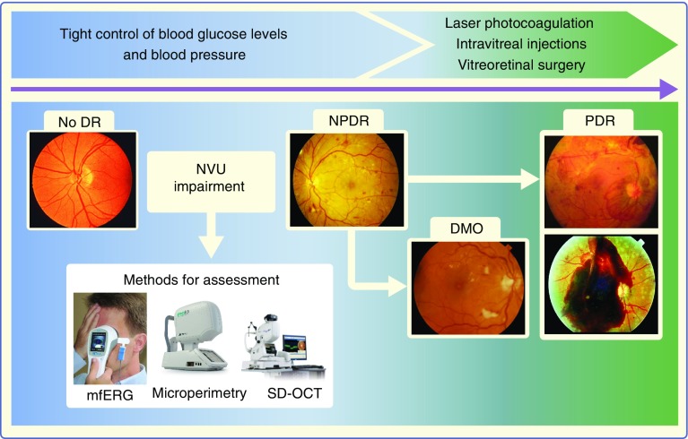
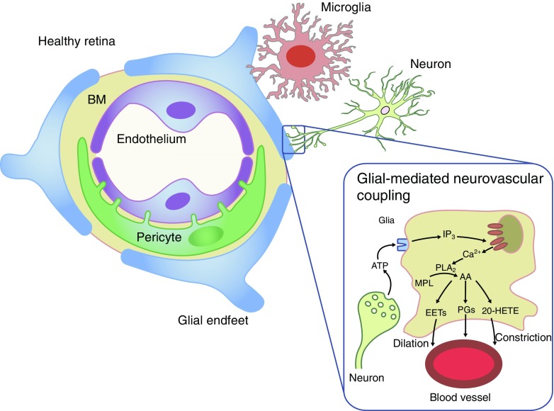
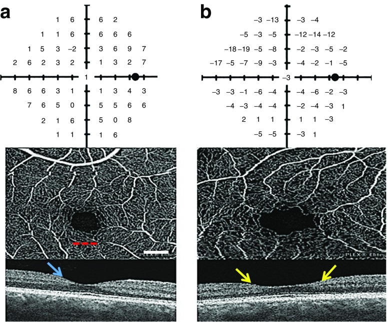
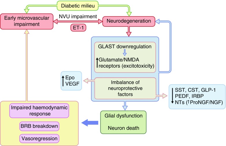
The concept of diabetic retinopathy as a microvascular disease has evolved, in that it is now considered a more complex diabetic complication in which neurodegeneration plays a significant role. In this article we provide a critical overview of the role of microvascular abnormalities and neurodegeneration in the pathogenesis of diabetic retinopathy. A special emphasis is placed on the pathophysiology of the neurovascular unit (NVU), including the contributions of microvascular and neural elements. The potential mechanisms linking retinal neurodegeneration and early microvascular impairment, and the effects of neuroprotective drugs are summarised. Additionally, we discuss how the assessment of retinal neurodegeneration could be an important index of cognitive status, thus helping to identify individuals at risk of dementia, which will impact on current procedures for diabetes management. We conclude that glial, neural and microvascular dysfunction are interdependent and essential for the development of diabetic retinopathy. Despite this intricate relationship, retinal neurodegeneration is a critical endpoint and neuroprotection, itself, can be considered a therapeutic target, independently of its potential impact on microvascular disease. In addition, interventional studies targeting pathogenic pathways that impact the NVU are needed. Findings from these studies will be crucial, not only for increasing our understanding of diabetic retinopathy, but also to help to implement a timely and efficient personalised medicine approach for treating this diabetic complication.

Keywords: Diabetic retinopathy; Microvascular impairment; Neurodegeneration; Neuroprotection; Neurovascular unit; Personalised medicine; Review.


Conflict of interest statement

The authors declare that there is no duality of interest associated with this manuscript.

Figures

Fig. 1
Fig. 1
Natural history of diabetic retinopathy, based on retinal microvascular disease progression, and current treatment options. NVU impairment is an early event in the pathogenesis of diabetic retinopathy that can be assessed by functional (i.e. mfERG [with or without flickering] and microperimetry) or morphological (i.e. SD-OCT) analysis. DR, diabetic retinopathy; mfERG, multifocal electroretinogram; NPDR, non-proliferative diabetic retinopathy; PDR, proliferative diabetic retinopathy; SD-OCT, spectral domain OCT. Schematic adapted from Simó and Hernández [73] by permission from BMJ Publishing Group Limited. mfERG image, distributed under the terms of the Creative Commons Attribution-Share Alike 4.0 International License (https://creativecommons.org/licenses/by-sa/4.0/); microperimetry image, used with permission from CenterVue SpA; SD-OCT image, 3D OCT-2000, used by permission of Topcon GB Ltd. This figure is available as part of a downloadable slideset
Fig. 2
Fig. 2
Composition of the retinal NVU. The NVU consists of vascular elements (endothelial cells, pericytes), the basement membrane (BM), glial cells (Müller cells, astrocytes), microglia and neurons. Glial-mediated neurovascular coupling is schematically represented. Synaptic release of ATP from neurons stimulates purinergic receptors on glial cells, leading to the production of inositol trisphosphate (IP3) and the release of Ca2+ from internal stores. Ca2+ activates phospholipase A2 (PLA2), which converts membrane phospholipids (MPL) to arachidonic acid (AA), which is subsequently metabolised to the vasodilators prostaglandin E2 (PGs) and epoxyeicosatrienoic acids (EETs), and to the vasoconstrictor 20-hydroxy-eicosatetraenoic acid (20-HETE) [23]. Interestingly, glial-induced vasodilating prostanoids are active at low NO concentrations, whereas vasoconstricting prostanoids are predominant at higher NO concentrations [24]. Healthy retina adapted from an illustration by R. Davidowitz in Duh et al [21], distributed under the terms of the Creative Commons Attribution 4.0 International License (http://creativecommons.org/licenses/by/4.0/), which permits unrestricted use, distribution, and reproduction in any medium. Glial-mediated neurovascular coupling illustration adapted from a drawing by A. Mishra in a review by Eric Newman [22], © SAGE Publications. This figure is available as part of a downloadable slideset
Fig. 3
Fig. 3
Frequency-doubling perimetry field tests with corresponding OCT angiograms and macular OCT (a) for a healthy 58 year old woman with 20/16 visual acuity and (b) for a 62 year old woman with an 18 year history of type 2 diabetes, 20/25 visual acuity and gastroparesis, but with a normal clinical examination and fluorescein angiogram (data not shown). The individual with diabetes has reduced frequency-doubling perimetry (FDP) sensitivity confirmed by repeat testing, an enlarged and irregular foveal avascular zone, a wide fovea and generalised inner retinal thinning compared with the control individual (T. W. Gardner and A. Omari, unpublished data). The red dotted line underlies the foveal avascular zone. The normal foveal depression is denoted by the blue arrow. The two yellow arrows denote inner retinal thinning. Scale bar, 0.5 mm. This figure is available as part of a downloadable slideset
Fig. 4
Fig. 4
Main features of neurodegeneration: glial activation (also known as reactive gliosis) and neural apoptosis. (a, c) Glial activation (green), assessed by analysis of glial fibrillary acidic protein (GFAP) expression, and (b, d) neural apoptosis, analysed using TUNEL assay in retinas from (a, b) an experimental model of type 2 diabetes (db/db mouse) and a control (db/+) mouse and (c, d) human diabetic and non-diabetic donors. (e) Image obtained by transmission electron microscopy showing DNA fragmentation in photoreceptors in db/db mice, which is characteristic of the apoptotic process. The nuclei of cells are stained in blue. The arrows indicate glial activation (a, c) and apoptotic cells (b, d). (a–d) Scale bar, 20 μm; (e) scale bar, 5μm. GCL, ganglion cell layer; INL, inner nuclear layer; ONL, outer nuclear layer; T2D, type 2 diabetes. (a, b, d, e), images from R. Simó’s laboratory, not previously published; (c) Adapted from Carrasco et al [86], distributed under the terms of the Creative Commons Attribution-NonCommercial-NoDerivatives License 3.0 (http://creativecommons.org/licenses/by-nc-nd/3.0/). This figure is available as part of a downloadable slideset
Fig. 5
Fig. 5
Main mechanisms involved in the link between retinal neurodegeneration and microvascular impairment in diabetes. Impaired cell–cell interaction within the NVU is critical in the pathogenesis of early stages of diabetic retinopathy. For example, ET-1 (levels of which are altered in diabetes) has a significant role in inducing both vasoconstriction and neurodegeneration via activation of the endothelin receptors ETA and ETB, respectively. In addition, increased extracellular concentrations of glutamate, owing to downregulation of the glutamate aspartate transporter (GLAST) in those with diabetes, results in excitotoxicity and neuron death. The subsequent progressive imbalance between neuroprotective factors (somatostatin, cortistatin, glucagon-like peptide 1, PEDF etc) is a major factor in neural apoptosis and glial activation in diabetes. In terms of vascular impairment, this leads to an altered haemodynamic response, BRB breakdown and vasoregression. CST, cortistatin; Epo, erythropoietin; IRBP, interphotoreceptor retinoid-binding protein; NGF, nerve growth factor; NMDA, N-methyl-d-aspartate; NT, neurotrophin; ProNGF, nerve growth factor precursor; SST, somatostatin. Adapted from Simó and Hernández [4], with permission from Elsevier. This figure is available as part of a downloadable slideset

References

    1. Yau JW, Rogers SL, Kawasaki R, et al. Global prevalence and major risk factors of diabetic retinopathy. Diabetes Care. 2012;35:556–564. doi: 10.2337/dc11-1909. - DOI - PMC - PubMed
    1. Leasher JL, Bourne RR, Flaxman SR, et al. Global estimates on the number of people blind or visually impaired by diabetic retinopathy: a meta-analysis from 1990 to 2010. Diabetes Care. 2016;39:1643–1649. doi: 10.2337/dc15-2171. - DOI - PubMed
    1. Abcouwer SF, Gardner TW. Diabetic retinopathy: loss of neuroretinal adaptation to the diabetic metabolic environment. Ann N Y Acad Sci. 2014;1311:174–190. doi: 10.1111/nyas.12412. - DOI - PMC - PubMed
    1. Simó R, Hernández C. Neurodegeneration in the diabetic eye: new insights and therapeutic perspectives. Trends Endocrinol Metab. 2014;25:23–33. doi: 10.1016/j.tem.2013.09.005. - DOI - PubMed
    1. Simó R, Hernández C. Novel approaches for treating diabetic retinopathy based on recent pathogenic evidence. Prog Retin Eye Res. 2015;48:160–180. doi: 10.1016/j.preteyeres.2015.04.003. - DOI - PubMed

Publication types

MeSH terms

Substances

LinkOut - more resources