

Neuroimaging in the Kleine-Levin Syndrome
- PMID: 30030664
- PMCID: PMC6061192
- DOI: 10.1007/s11910-018-0866-y
Neuroimaging in the Kleine-Levin Syndrome
Abstract
Purpose of review: The purpose was to review the most recent literature on neuroimaging in the Kleine-Levin syndrome (KLS). We aimed to investigate if frontotemporal and thalamic dysfunction are key KLS signatures, and if recent research indicates other brain networks of interest that elucidate KLS symptomatology and aetiology.
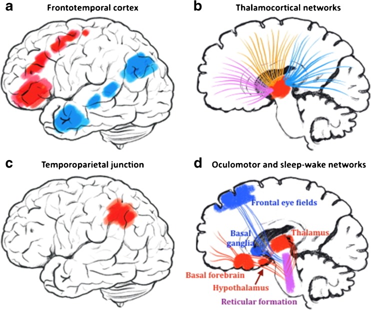
Recent findings: In a comprehensive literature search, we found 12 original articles published 2013-2018. Most studies report deviations related to cerebral perfusion, glucose metabolism, or blood-oxygen-level-dependent responses in frontotemporal areas and/or the thalamus. Studies also report dysfunction in the temporoparietal junction and the oculomotor network that also were related to clinical parameters. We discuss these findings based on recent research on thalamocortical networks and brain stem white matter tracts. The hypothesis of frontotemporal and thalamic involvement in KLS was confirmed, and additional findings in the temporoparietal junction and the oculomotor system suggest a broader network involvement, which can be investigated by future high-resolution and multimodal imaging.
Keywords: Diffusion weighted imaging (DWI); Functional magnetic resonance imaging (fMRI); Kleine-Levin syndrome (KLS); Magnetic resonance spectroscopy (MRS); Positron emission tomography (PET); Single photon emission computed tomography (SPECT).
Conflict of interest statement
Conflict of Interest
Maria Engström, Francesco Latini, and Anne-Marie Landtblom each declare no potential conflicts of interest.
Human and Animal Rights and Informed Consent
This article does not contain any studies with human or animal subjects performed by any of the authors.
Figures
References
-
- Engström M. Neuroimaging in Kleine-Levin syndrome. In: Thorpy M, Nofzinger E, Maquet P, editors. Neuroimaging of sleep and sleep disorders: Cambridge University Press; 2013.
Publication types
MeSH terms
LinkOut - more resources
Full Text Sources
Other Literature Sources
Research Materials
