. 2019 Feb;74(2):236-245.
doi: 10.1111/all.13567. Epub 2018 Nov 20.

The soluble isoform of human FcɛRI is an endogenous inhibitor of IgE-mediated mast cell responses

Affiliations

The soluble isoform of human FcɛRI is an endogenous inhibitor of IgE-mediated mast cell responses

S Moñino-Romero et al. Allergy. 2019 Feb.

Abstract

Background: The soluble isoform of FcɛRI, the high-affinity IgE receptor (sFcεRI), is a protein of the IgE network with poorly defined functions.

Objective: To define cellular sources and signals that result in the production of human sFcεRI and study its in vivo functions.

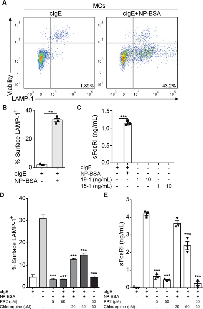
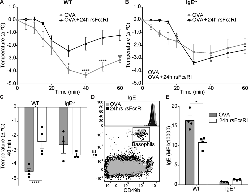
Methods: FcεRI-transfected human cell lines (MelJuso), human monocyte-derived dendritic cells (moDCs), and murine bone marrow-derived mast cells (MC) were stimulated by FcεRI cross-linking and release of sFcεRI was analyzed (ELISA, Western Blot). Lysosomal-associated membrane protein 1 degranulation assays and human basophil activation tests (BATs) were used to study IgE-dependent activation. Recombinant sFcεRI (rsFcεRI) was used to assess its role in murine models of anaphylaxis with WT (wild-type) and IgE-/- (IgE-deficient) mice.

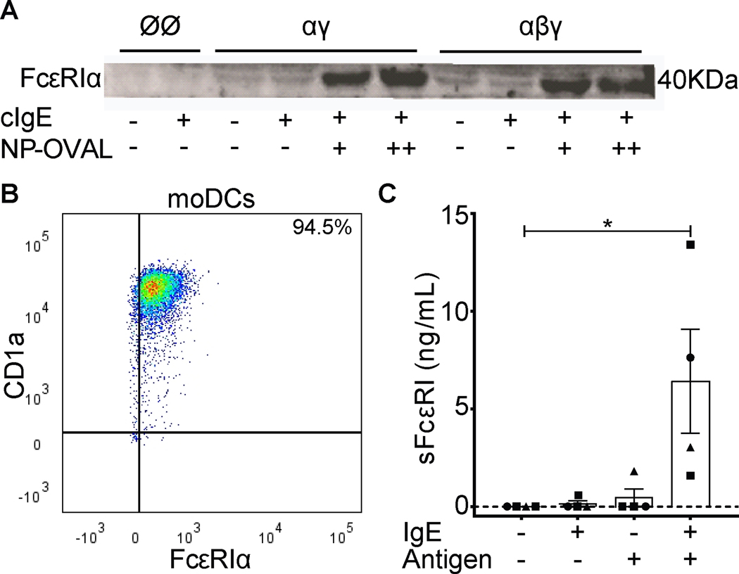
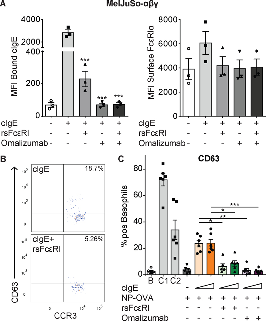
Results: Antigen-specific cross-linking of IgE-loaded FcɛRI on MelJuso cells that express the trimeric or tetrameric receptor isoform induced the production of sFcεRI. Using MCs and moDCs, we confirmed that IgE/FcɛRI activation induces sFcɛRI release. We demonstrated that generation of sFcɛRI requires Src phosphorylation and endo/lysosomal acidification. In experimental mouse models, sFcɛRI diminishes the severity of IgE-mediated anaphylaxis. BATs confirmed that, comparable to the anti-IgE monoclonal antibody omalizumab, sFcɛRI is an inhibitor of the human innate IgE effector axis, implying that sFcɛRI and omalizumab potentially inhibit each other in vivo.

Conclusion: sFcɛRI is produced after antigen-specific IgE/FcɛRI-mediated activation signals and functions as an endogenous inhibitor of IgE loading to FcɛRI and IgE-mediated activation. Our results imply, therefore, that sFcɛRI contributes to a negative regulatory feedback loop that aims at preventing overshooting responses after IgE-mediated immune activation.

Keywords: FcεRI; IgE receptor; allergy; mast cell; omalizumab.


Conflict of interest statement

Conflicts of interest

The authors confirm that there are no known conflicts of interest associated with this publication and there has been no significant financial support for this work that could have influenced its outcome.

Figures

Figure 1.
Figure 1.. FcεRI-crosslinking on moDCs induces sFcεRI production.
Panel A shows detection of sFcεRI by Western Blot analysis from MelJusoØØ/-αγ/-αβγ cell cultures. Panel B shows a representative dot plot of mature monocyte-derived DCs. Panel C shows detection sFcεRI by ELISA analysis from monocyte-derived DCs given by mean ± SEM. cIgE (5–10 μg/mL) and NP-OVA (50–100 μg/mL). Individual points represent each donor where squares are males, circles females and triangle unknown. Kruskal-Wallis test plus Dunn’s multiple correction was performed where *p<0.05.
Figure 2.
Figure 2.. Src kinase activation and endo/lysosomal acidification are required for sFcεRI production.
Detection of surface LAMP-1 expression on bone marrow MCs by flow cytometry (A, B, D) and sFcεRI in supernatants by ELISA (C, E). Panel A shows representative dot plots of MCs from LAMP-1 degranulation assays. Panels B and D show the quantification of percentage of surface LAMP-1+ cells. Panels C and E show total sFcεRI levels from supernatant of MCs cultures. MCs were loaded overnight with cIgE (500 ng/mL) and added NP-BSA (100 ng/mL) for 10 minutes for LAMP-1 degranulation assays. PP2 and chloroquine were added for 30 min prior to NP-BSA. In parallel, MCs were loaded overnight with cIgE and added NP-BSA, 19–1, or 15–1 for 2 (19–1 and 15–1) or 24 hours before supernatants were harvested. Graphs show mean ± SEM where paired t-test (B) or 1way ANOVA tests plus Tukey’s multiple correction (C-E) was performed; where **p<0.01 and ***p<0.001 compared to cells incubated with cIgE and NP-BSA.
Figure 3.
Figure 3.. sFcεRI blocks cell surface cIgE binding and basophil activation.
Detection of bound cIgE (A, left) and FcεRIα (A, right) on MelJuso-αβγ (A) cells by flow cytometry (sFcεRI (25 nM) and omalizumab (0.27 μM)). Graphs show mean ± SEM. Individual points represent means of independent experiments (n=3). Detection of surface CD63 and CCR3 on basophils by flow cytometry. Panel B shows representative dot plots for basophils loaded with cIgE in presence or absence of rsFcεRI. Panel C shows the percentage of positive CD63 basophils stimulated with NP-OVA (2.85 μM) and sFcεRI (0.52 μM) or omalizumab (0.8 μM). Graphs show mean ± SEM. Individual points represent each donor (n=6). 1way ANOVA tests plus Tukey’s multiple correction were performed where *p<0.05, **p<0.01, and ***p<0.001, compared to cells loaded with cIgE. B: background; C1: anti-FcεRI mAb; C2: fMLP.
Figure 4.
Figure 4.. sFcεRI prevents systemic sensitization and improves recovery.
Analysis of core body temperature from passively (A-C) and systemically (D, E) sensitized mice. Panel A shows the passive sensitization and challenge strategy. Panel B shows the time course of core body temperature drop in sensitized mice that were challenged in the absence or presence of rsFcεRI. Representative experiment, average temperature of 3–6 mice per time point. Panel C shows the composite graph of temperature drops at the 20 min time point following DNP-OVA challenge, from three independent experiments (n= 3, total of 19 mice). Panel D shows the Th2-type sensitization and challenge strategy. Panel E shows the time course of body temperature drop in sensitized mice that were challenged with OVA in absence or presence of rsFcεRI 24 hours before challenge. rsFcεRI (3 μg), IgE-anti-DNP (5 μg), OVA/Alum (100 μg), and OVA (100–500 μg). Graphs show mean ± SEM with n=3–6/group where 1way ANOVA test and Tukey’s multiple correction (C), or 2way ANOVA tests and Tukey’s (B) or Bonferroni’s (E) multiple correction were performed; *p<0.05; **p<0.01; ***p<0.001, and ****p<0.0001. * represents statistics between IgE and dIgE (B), and PBS and OVA (E) groups, *§ represents statistics between IgE-anti-DNP and IgE-anti-DNP + rsFcεRI (B), and OVA and OVA+24h rsFcεRI (E) groups.
Figure 5.
Figure 5.. Recombinant sFcεRI prevents anaphylaxis and reduces FcεRI bound IgE on the surface of basophils.
Analysis of rectal temperature from systemically sensitized mice (A-C). Panels A and B compare the effect of rsFcεRI treatment in a time course of core temperature drop in WT (A) or IgE−/− (B) mice. Panel C shows the maximum temperature drop of WT and IgE−/− mice 40 min after OVA challenge. Panel D shows a representative histogram and a dot plot of peripheral blood basophils from WT mice with and without rsFcεRI treatment. Panel E shows the MFI of surface bound IgE on basophils with and without rsFcεRI treatment. rsFcεRI (3 μg), OVA/Alum (100 μg), and OVA (500 μg). Graphs show mean ± SEM with n=3–4/group where 2way ANOVA tests and Tukey’s multiple correction were performed; *p<0.05 and ****p<0.0001.

References

    1. Kraft S, Kinet JP. New developments in FcepsilonRI regulation, function and inhibition. Nature reviews Immunology. 2007;7(5):365–78. - PubMed
    1. von Bubnoff D, Novak N, Kraft S, Bieber T. The central role of FcepsilonRI in allergy. Clinical and experimental dermatology. 2003;28(2):184–7. - PubMed
    1. Gould HJ, Sutton BJ. IgE in allergy and asthma today. Nature reviews Immunology. 2008;8(3):205–17. - PubMed
    1. Hogan SP, Wang YH, Strait R, Finkelman FD. Food-induced anaphylaxis: mast cells as modulators of anaphylactic severity. Semin Immunopathol. 2012;34(5):643–53. - PMC - PubMed
    1. Reber LL, Hernandez JD, Galli SJ. The pathophysiology of anaphylaxis. J Allergy Clin Immunol. 2017;140(2):335–48. - PMC - PubMed

Publication types

MeSH terms