Review
. 2018 Dec:53:88-99.
doi: 10.1016/j.sbi.2018.06.004. Epub 2018 Jul 19.

Detecting and interpreting DNA methylation marks

Affiliations
Review

Detecting and interpreting DNA methylation marks

Ren Ren et al. Curr Opin Struct Biol. 2018 Dec.

Abstract

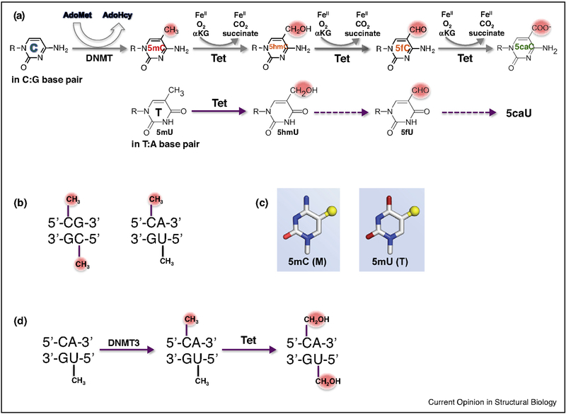
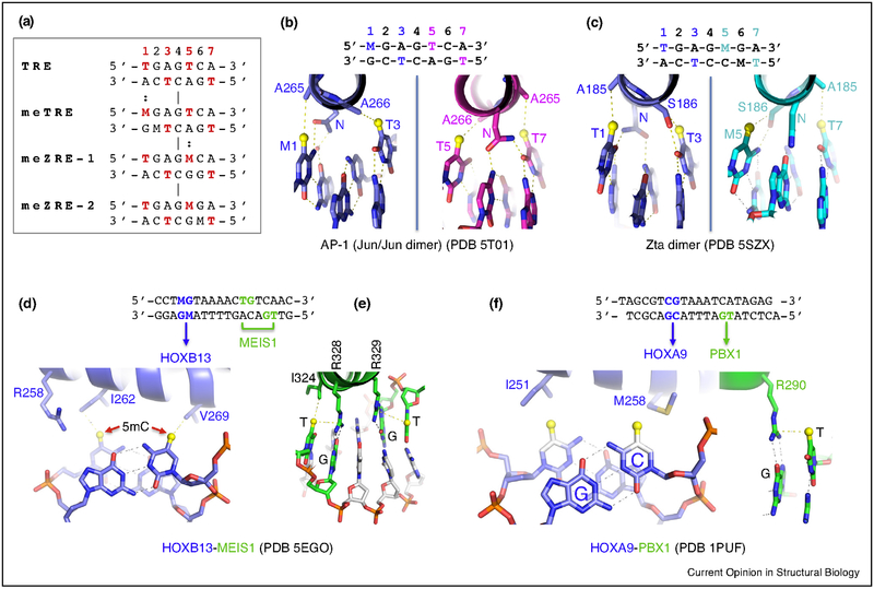
The generation, alteration, recognition, and erasure of epigenetic modifications of DNA are fundamental to controlling gene expression in mammals. These covalent DNA modifications include cytosine methylation by AdoMet-dependent methyltransferases and 5-methylcytosine oxidation by Fe(II)-dependent and α-ketoglutarate-dependent dioxygenases. Sequence-specific transcription factors are responsible for interpreting the modification status of specific regions of chromatin. This review focuses on recent developments in characterizing the functional and structural links between the modification status of two DNA bases: 5-methylcytosine and 5-methyluracil (thymine).


Conflict of interest statement

Conflict of interest statement

The authors declare no conflict of interest.

Figures

Figure 1
Figure 1
Methylation and oxidation in 5′-CpG-3′ and 5′-CpA-3′ dinucleotides. (a) DNA methyltransferases (DNMTs) convert C to 5-methylC (5mC). Tet dioxygenases then convert 5mC to 5-hydroxymethylC (5hmC), 5-formylC (5fC), and 5-carboxyC (5caC) in three consecutive Fe(II)-dependent and α-ketoglutarate-dependent oxidation reactions, without release of formaldehyde (Top). Tet dioxygenases can also convert thymine (5mU) to 5hmU, and potentially to 5fU and 5caU (Bottom). (b) CpG/CpG can be methylated on both strands; while TpG/CpA is intrinsically methylated on one strand (T, 5mU), and the C on the other strand may be methylated as well. (c) 5mC and 5mU (T) contain a methyl group (yellow sphere) in spatially equivalent positions at C5. (d) Dnmt3A and Dnmt3B can methylate cytosine in the context of a CpA/TpG dinucleotide. Tet dioxygenases can oxidize both 5mC and T (5mU).
Figure 2
Figure 2
Examples of methyl-Arg-Gua recognition and 5caC recognition. (a) MBD domain of MeCP2. PDB codes are given, DNA recognition sequences are shown with M = 5mC, and key interactions of each DNA strand are shown above and below the recognition sequence. (b) Kaiso (ZF), (c) Zfp57 (ZF), (d) Klf4 (ZF), (e) Egr1 (ZF) and (f) WT1 (ZF). (g) Effects of binding of DNA with 5caC against Egr1 and WT1. (h) WT1 (Q369) recognition of DNA with 5caC. (i) The p53 proteins recognize TpG, which can be replaced by 5mCpG. (j) Klf4 mutant (D446) interacts with unmodified cytosine. (k) In CTCF (ZF), D451 and E362 recognize, respectively, unmodified cytosine at position 2 and 5mC at position 12. (l) Effects of methylation at specific sites on binding by CTCF. (m) MAX (bHLH) recognition of 5caC. (n) Pol II recognition of 5caC. (o) Tet3 CXXC domain recognition of 5caC.
Figure 3
Figure 3
Examples of methyl group recognition via van der Waals contacts. (a) Aligned DNA response elements with spatially equivalent methyl groups from T (5mU) or M (5mC). (b,c) Recognition of the four spatially constrained methyl groups of meTRE by Jun/Jun dimer (bZIP) or meZRE-2 by Zta dimer (bZIP). (d) Recognition of the two methyl groups of 5mCpG duplex by HOXB13 (homeodomain). (e) Homeodomain protein MEIS1 recognizes TpG via methyl–Arg–Gua triad. (f) HOXA9–PBX1 in complex with DNA. Both proteins could have preferential binding of methylated DNA. A methyl group (in yellow sphere) is modeled onto unmodified cytosine.

References

    1. Kriaucionis S, Heintz N: The nuclear DNA base 5-hydroxymethylcytosine is present in Purkinje neurons and the brain. Science 2009, 324:929–930. - PMC - PubMed
    1. Tahiliani M, Koh KP, Shen Y, Pastor WA, Bandukwala H, Brudno Y, Agarwal S, Iyer LM, Liu DR, Aravind L et al.: Conversion of 5-methylcytosine to 5-hydroxymethylcytosine in mammalian DNA by MLL partner TET1. Science 2009, 324:930–935. - PMC - PubMed
    1. Ito S, Shen L, Dai Q, Wu SC, Collins LB, Swenberg JA, He C, Zhang Y: Tet proteins can convert 5-methylcytosine to 5-formylcytosine and 5-carboxylcytosine. Science 2011, 333:1300–1303. - PMC - PubMed
    1. He YF, Li BZ, Li Z, Liu P, Wang Y, Tang Q, Ding J, Jia Y, Chen Z, Li L et al.: Tet-mediated formation of 5-carboxylcytosine and its excision by TDG in mammalian DNA. Science 2011, 333:1303–1307. - PMC - PubMed
    1. Pfaffeneder T, Hackner B, Truss M, Munzel M, Muller M, Deiml CA, Hagemeier C, Carell T: The discovery of 5-formylcytosine in embryonic stem cell DNA. Angew Chem Int Ed Engl 2011, 50:7008–7012. - PubMed

Publication types

LinkOut - more resources