. 2018 Jul 21;37(1):162.
doi: 10.1186/s13046-018-0825-0.

Exosomal TRIM3 is a novel marker and therapy target for gastric cancer

Affiliations

Exosomal TRIM3 is a novel marker and therapy target for gastric cancer

Hailong Fu et al. J Exp Clin Cancer Res. .

Abstract

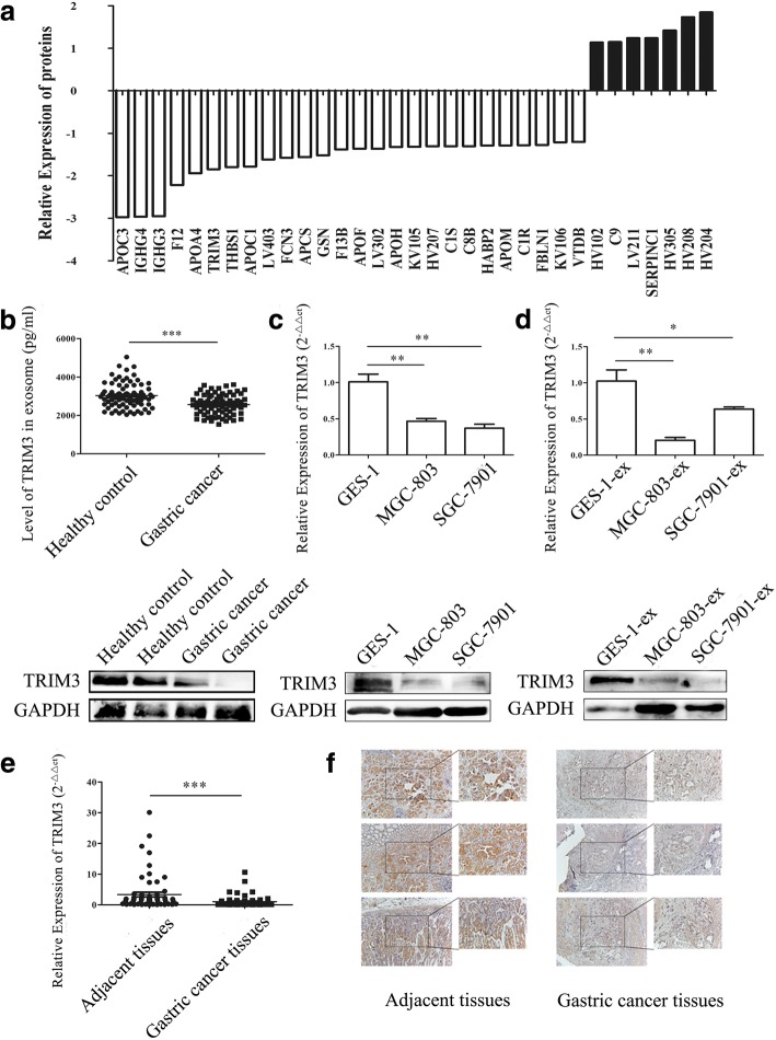
Background: Exosomes are critically involved in cancer development and progression. The exosomal contents have been suggested as ideal cancer biomarkers. In this study, we investigated the expression of exosomal proteins in the serum of gastric cancer patients and their roles in gastric cancer.

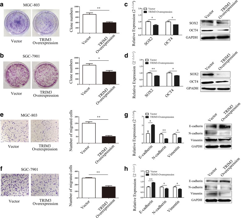
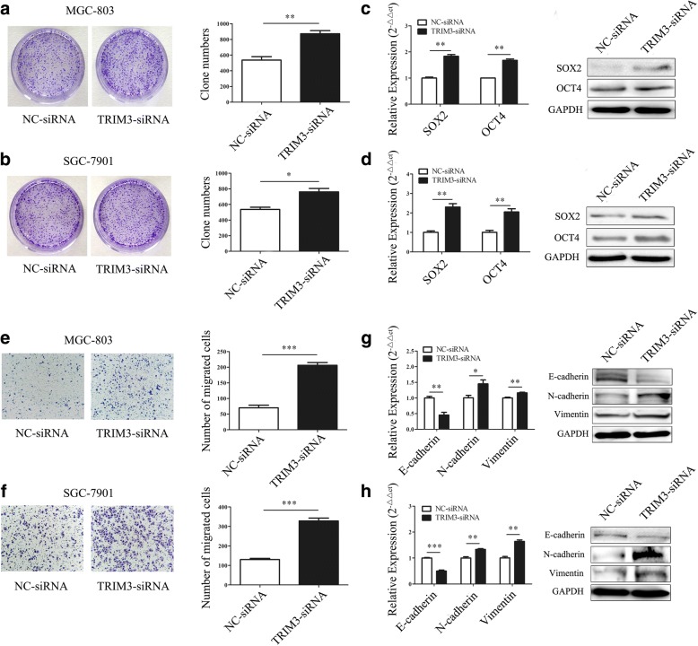
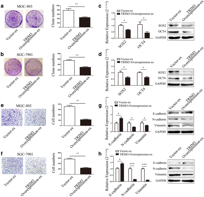
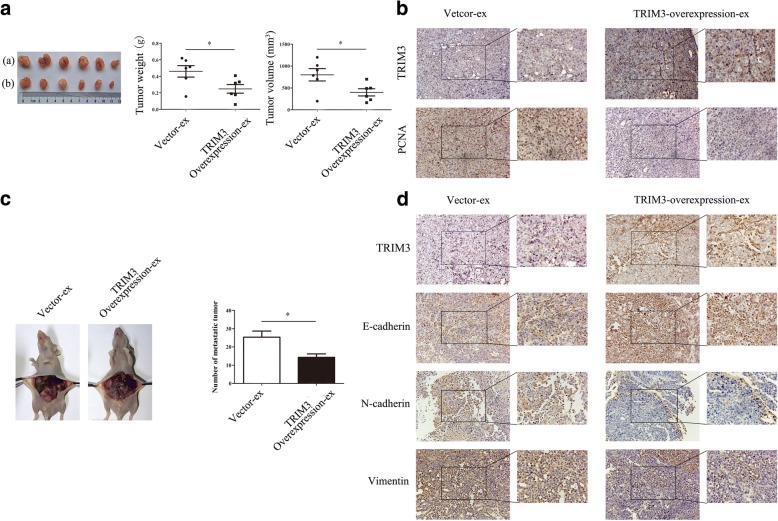
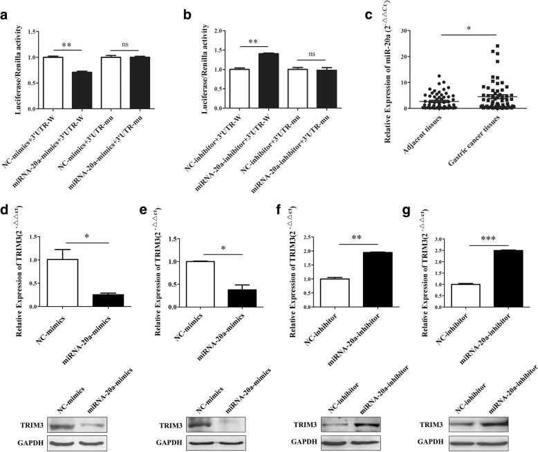
Methods: The proteomic profile of exosomes from the serum of gastric cancer patients was detected by using LC-MS/MS. The expression of TRIM3 in exosomes from the serum of gastric cancer patients and healthy controls was assessed by ELISA and western blot. Immunohistochemistry was used to detect TRIM3 expression in gastric cancer tissues and their matching adjacent tissues. The growth and migration abilities of gastric cancer cells with TRIM3 overexpression or knockdown in vitro were evaluated by colony formation assay and transwell migration assay. The effects of TRIM3 overexpression or knockdown on gastric cancer growth and metastasis in vivo were investigated by using subcutaneous xenograft tumor and peritoneal metastasis mouse model. The effects of TRIM3-overexpressing exosomes on gastric cancer growth and metastasis in vitro and in vivo were also evaluated.

Results: We found that the expression levels of TRIM3 mRNA and protein were decreased in gastric cancer tissues compared to the matched control tissues. In addition, the levels of TRIM3 protein in the serum exosomes of gastric cancer patients were lower than that in healthy controls. We demonstrated that TRIM3 overexpression reduced while TRIM3 knockdown promoted the growth and metastasis of gastric cancer in vitro and in vivo through the regulation of stem cell factors and EMT regulators. Moreover, exosomes-mediated delivery of TRIM3 protein could suppress gastric cancer growth and metastasis in vitro and in vivo.

Conclusions: Taken together, our findings suggest that exosomal TRIM3 may serve as a biomarker for gastric cancer diagnosis and the delivery of TRIM3 by exosomes may provide a new avenue for gastric cancer therapy.

Keywords: Diagnosis; Exosomes; Gastric cancer; Progression; TRIM3; Therapy.


Conflict of interest statement

Ethics approval and consent to participate

This study was approved by the ethics committee of Jiangsu University (2012258), and written informed consent was obtained from all patients.

Consent for publication

Not applicable.

Competing interests

The authors declare that they have no competing interests.

Publisher’s Note

Springer Nature remains neutral with regard to jurisdictional claims in published maps and institutional affiliations.

Figures

Fig. 1
Fig. 1
The characterization of exosomes from the serum of gastric cancer patients and the culture supernatant of gastric cancer cell lines. a, The size and morphology of exosomes from the serum of gastric cancer patients and healthy controls ((a), Nanosight analyses; (b), TEM analyses). b, The concentration of exosomes from the serum of gastric cancer patients and healthy controls. **P < 0.01. c, The mean diameter of exosomes from the serum of gastric cancer patients and healthy controls. d, The size and morphology of exosomes from the culture supernatants of gastric epithelial cells and gastric cancer cells ((a), Nanosight analyses; (b), TEM analyses). e, The concentration of exosomes from the culture supernatants of gastric epithelial cells and gastric cancer cells, **P < 0.01. f, The mean diameter of exosomes from the culture supernatants of gastric epithelial cells and gastric cancer cells
Fig. 2
Fig. 2
The proteomic analysis of TRIM3 expression in exosomes. a, LC-MS/MS analyses of the protein profiles of exosomes from the serum of gastric cancer patients and healthy controls. b, ELISA and western blot analyses of the expression of TRIM3 protein in exosomes from the serum of gastric cancer patients and healthy controls, ***P < 0.001. c, The expression of TRIM3 gene in gastric epithelial cells and gastric cancer cells was examined by using qRT-PCR and western blot. **P < 0.01. d, The expression of TRIM3 gene in exosomes from the culture supernatants of gastric epithelial cells and gastric cancer cells was detected by using qRT-PCR and western blot. *P < 0.05, **P < 0.01. e, The expression of TRIM3 gene in gastric cancer tissues and adjacent control tissues was detected by using qRT-PCR. ***P < 0.001. f, The expression of TRIM3 protein in gastric cancer tissues and adjacent control tissues was detected by using immunohistochemistry
Fig. 3
Fig. 3
TRIM3 overexpression inhibits gastric cancer cell proliferation and migration in vitro. a and b, TRIM3 overexpression inhibits the colony formation abilities of MGC-803 (a) and SGC-7901 (b) cells. *P < 0.05, **P < 0.01. c and d, The expression of SOX2 and OCT4 in gastric cancer cells transfected with TRIM3, c, MGC-803; d, SGC-7901. *P < 0.05, **P < 0.01. e and f, TRIM3 overexpression inhibits the migration of MGC-803 (e) and SGC-7901 (f) cells. Magnification, 100×; **P < 0.01. g and h, The expression of EMT associated genes in MGC-803 (g) and SGC-7901 (h) cells transfected with TRIM3. *P < 0.05, **P < 0.01
Fig. 4
Fig. 4
TRIM3 knockdown promotes the proliferation and migration of gastric cancer cells in vitro. a and b, TRIM3-siRNA promotes the colony formation abilities of MGC-803 (a) and SGC-7901 (b) cells. *P < 0.05, **P < 0.01. c and d, The expression of SOX2 and OCT4 in gastric cancer cells transfected with TRIM3-siRNA. (c) MGC-803; (d) SGC-7901. **P < 0.01. e and f, TRIM3-siRNA enhances the migration of MGC-803 (e) and SGC-7901 (f) cells. Magnification 100×; ***P < 0.001. g and h, The expression of EMT associated genes in MGC-803 (g) and SGC-7901 (h) cells transfected with TRIM3-siRNA. *P < 0.05, **P < 0.01, ***P < 0.001
Fig. 5
Fig. 5
TRIM3 overexpression inhibits gastric cancer growth and metastasis in vivo. a, The weight and volumes of tumors in control and TRIM3 overexpression groups. ((a). TRIM3-Vector; (b), TRIM3 overexpression). *P < 0.05, ** P < 0.01. b, The expression of TRIM3 and PCNA in subcutaneous tumor tissues was examined by using immunohistochemistry. Magnification, 200× (left panel); 400× (right panel). c, The number of metastatic tumor nodes in control and TRIM3 overexpression groups. **P < 0.01. d, The expression of TRIM3 and EMT associated proteins in the metastatic tumor nodes was detected by using immunohistochemistry. Magnification, 200× (left panel); 400× (right panel). e, The weight and volume of tumors in control and TRIM3-siRNA groups. *P < 0.01; **P < 0.05. f, The expression of TRIM3 and PCNA in subcutaneous tumor tissues was detected by using immunohistochemistry. Magnification, 200× (left panel); 400× (right panel). g, The number of metastatic tumor nodes in control and TRIM3-siRNA groups. *P < 0.05. h, The expression of TRIM3 and EMT associated proteins in the metastatic tumor nodes was detected by using immunohistochemistry. Magnification, 200× (left panel); 400× (right panel)
Fig. 6
Fig. 6
Exosomal TRIM3 inhibits the proliferation and migration of gastric cancer cells in vitro. a and b, The effects of TRIM3-overexpressing exosomes on the colony formation abilities of MGC-803 and SGC-7901 cells. **P < 0.01. c and d, The expression of SOX2 and OCT4 genes and proteins in MGC-803 (c) and SGC-7901 (d) cells treated with 50 μg TRIM3-overexpressing exosomes. *P < 0.05. e and f, The effects of TRIM3-overexpressing exosomes on the migration of MGC-803 (e) and SGC-7901 (f) cells. Magnification, 100×; *P < 0.05, **P < 0.01. g and h, The expression of EMT associated genes and proteins in MGC-803 (g) and SGC-7901 (h) cells treated with 50 μg TRIM3-overexpressing exosomes. *P < 0.05, ***P < 0.001
Fig. 7
Fig. 7
Exosomal TRIM3 inhibits tumor growth and metastasis in vivo. a, The effects of TRIM3-overexpressing exosomes on tumor growth in vivo. ((a), control exosomes group; (b), TRIM3-overexpressing exosome group). *P< 0.05. b, The expression of TRIM3 and PCNA in subcutaneous tumor tissues in control exosome and TRIM3-overexpressing exosome groups. Magnification, 200× (left panel); 400× (right panel). c, The effects of TRIM3-overexpressing exosomes on tumor metastasis in vivo. The number of metastatic tumor nodes in control exosomes and TRIM3-overexpressing exosome groups were compared. *P < 0.05. d, The expression of TRIM3 and EMT associated proteins in the metastatic tumor tissues in control exosome and TRIM3-overexpressing exosome groups. Magnification, 200× (left panel); 400× (right panel)
Fig. 8
Fig. 8
TRIM3 downregulation is associated with miR-20a in gastric cancer. a, miR-20a mimics suppressed the luciferase activity of the wild type TRIM3 3’UTR (WT) (*P < 0.01) in HEK-293 cells. b, miR-20a-inhibitor enhanced the luciferase activity of the wild type TRIM3 3’UTR (WT) (*P < 0.01) in HEK-293 cells. c, miR-20a expression in gastric cancer tissues and adjacent control tissues, *P < 0.05. d and e, The expression of TRIM3 gene in miR-20a-mimics transfected MGC-803(d) and SGC-7901(e) examined by qRT-PCR and western blot. *P < 0.05. f and g, The expression of TRIM3 gene in miR-20a-inhibitor transfected MGC-803(f) and SGC-7901(g) examined by qRT-PCR and western blot. **P < 0.01, ***P < 0.001

References

    1. Jemal A, Siegel R, Ward E, Murray T, Xu J, Smigal C, Thun MJ. Cancer statistics, 2006. CA Cancer J Clin. 2006;56(2):106–130. doi: 10.3322/canjclin.56.2.106. - DOI - PubMed
    1. Saka M, Morita S, Fukagawa T, Katai H. Present and future status of gastric cancer surgery. Jpn J Clin Oncol. 2011;41(3):307–313. doi: 10.1093/jjco/hyq240. - DOI - PubMed
    1. Boukouris S, Mathivanan S. Exosomes in bodily fluids are a highly stable resource of disease biomarkers. Proteomics Clin Appl. 2015;9(3–4):358–367. doi: 10.1002/prca.201400114. - DOI - PMC - PubMed
    1. Thery C, Ostrowski M, Segura E. Membrane vesicles as conveyors of immune responses. Nat Rev Immunol. 2009;9(8):581–593. doi: 10.1038/nri2567. - DOI - PubMed
    1. Kowal J, Tkach M, Thery C. Biogenesis and secretion of exosomes. Curr Opin Cell Biol. 2014;29:116–125. doi: 10.1016/j.ceb.2014.05.004. - DOI - PubMed