. 2018 Aug 6;46(3):344-359.e4.
doi: 10.1016/j.devcel.2018.06.019. Epub 2018 Jul 19.

Fish Scales Dictate the Pattern of Adult Skin Innervation and Vascularization

Affiliations

Fish Scales Dictate the Pattern of Adult Skin Innervation and Vascularization

Jeffrey P Rasmussen et al. Dev Cell. .

Abstract

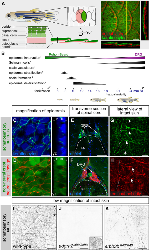
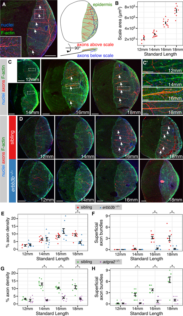
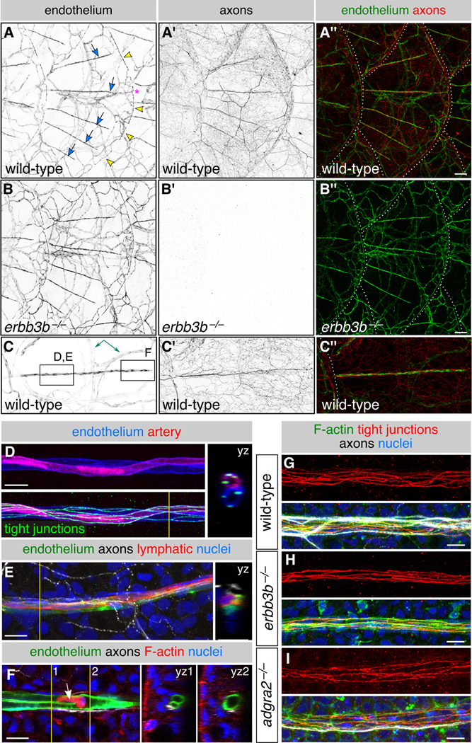
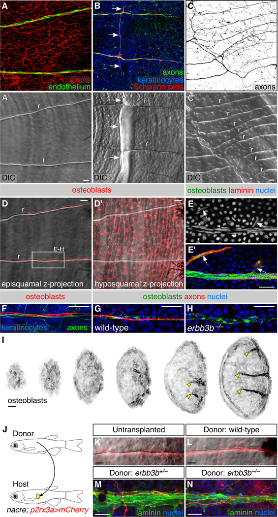
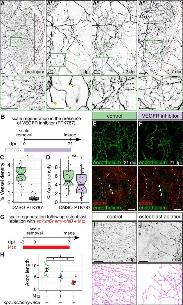
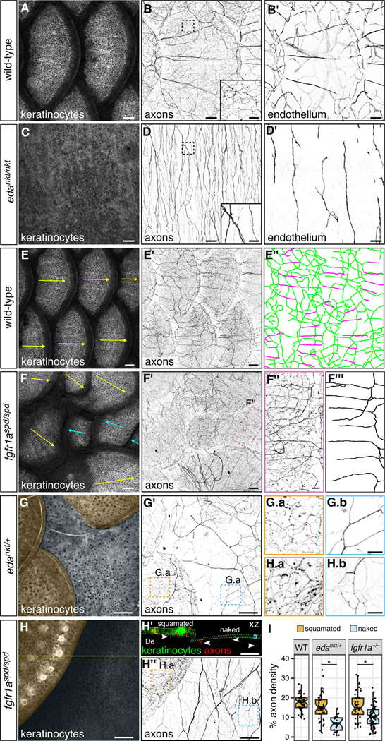
As animals mature from embryonic to adult stages, the skin grows and acquires specialized appendages, like hairs, feathers, and scales. How cutaneous blood vessels and sensory axons adapt to these dramatic changes is poorly understood. By characterizing skin maturation in zebrafish, we discovered that sensory axons are delivered to the adult epidermis in organized nerves patterned by features in bony scales. These nerves associate with blood vessels and osteoblasts above scales. Osteoblasts create paths in scales that independently guide nerves and blood vessels during both development and regeneration. By preventing scale regeneration and examining mutants lacking scales, we found that scales recruit, organize, and polarize axons and blood vessels to evenly distribute them in the skin. These studies uncover mechanisms for achieving comprehensive innervation and vascularization of the adult skin and suggest that scales coordinate a metamorphosis-like transformation of the skin with sensory axon and vascular remodeling.

Keywords: axon; dorsal root ganglia; metamorphosis; osteoblast; regeneration; scale; sensory nerve; skin; vasculature; zebrafish.


Figures

Figure 1.
Figure 1.. Adult epidermal innervation requires Erbb3b and Adgra2:
(A) Left, diagram of adult skin anatomy. Right, confocal projection and orthogonal views through the lateral trunk. Yellow lines, planes of orthogonal sections. In this and subsequent lateral views, posterior is to the right. (B) Timeline of trunk skin development with approximate timing of major morphogenetic events. See Figure S3A for timing of differentiation of club cells, a specialized cell type within the fish epidermis (Whitear, 1986). * shown in this study; † based on (Guzman et al., 2013); ‡ based on (Sire et al., 1997a). (C,D) Projections and orthogonal views through the epidermis of isolated scales. P, periderm; BC, basal cells. (E,F) Transverse sections through the mid-trunk. In this region, DRGs are located dorsal to the spinal cord (Weis, 1968). Double-headed arrows, DRG central axons within the spinal cord (sc). Dashed lines, spinal cord boundary. (G,H) Lateral views of intact adult trunk skin. Dashed lines, scale margins. Arrows, superficial axon bundles above scales. (I-K) Representative images of lateral trunk skin from adults of indicated genotypes. Inset in panel J, trigeminal sensory axons over the eye to confirm presence of transgene. Transgenes: (A) keratinocytes [Tg(krt4:EGFP)] (Rasmussen et al., 2015) and osteoblasts [Tg(sp7:mCherry-nfsB)] (Singh et al., 2012); (C) keratinocytes [Tg(actb2:BswitchR)] (Kobayashi et al., 2014); (C,E,G) somatosensory neurons [Tg(p2rx3b:EGFP)] (Kucenas et al., 2006); (D,F,H) Tg(−28.5Sox10:Cre);Tg(ubb:GswitchR) (Kague et al., 2012; Mosimann et al., 2011); (I-K) somatosensory axons Tg(p2rx3a>mCherry) (Palanca et al., 2013). Staining: (E,F) nuclei (DAPI). Scale bars, 100 μm (A,E-H), 10 μm (C,D) and 250 μm (I-K). See also Figures S1 and S2.
Figure 2.
Figure 2.. Remodeling of skin innervation during juvenile development:
(A) Adult scale removed from animal and immunostained for indicated structures. Note that the posterior region is covered by epidermis (green). (B) Measurements of scale size during late juvenile stages. n=8-12 scales/stage. (C) Representative scales from juvenile animals immunostained at the indicated stages. Magnifications of epidermis are shown in C’. Note that elongated tracts form before nerves enter the scale. (D) Representative scales from juvenile erbb3b−/− and sibling controls. Note that in erbb3b−/− scales axon bundles never form, and innervation is lost by 18 mm SL. Arrows in panels A, C and D indicate superficial axon bundles. (E-H) Quantification of axon density near the skin (E,G) and superficial axon bundles (F,H) based on acTubulin staining. n=4-12 scales/sample (mean sample size = 9.7). Black bars, mean ± SEM. *, p<0.01, Wilcoxon rank sum test. Staining: (A,C,D) F-actin (phalloidin), axons (acTubulin) and nuclei (DAPI). Scale bars, 250 μm (A), 100 μm (C,D) and 25 μm (C’). See also Figure S3.
Figure 3.
Figure 3.. DRG axons form nerves at the surface of scales:
(A-C) Projections through a single superficial scale nerve in an adult. Arrow, axon bundle. Arrowheads, axon free endings. Insets, orthogonal views of nerve; outer surface to left. (D) TEM of scale epidermis. Arrowheads, axon free endings ensheathed by keratinocyte membranes. (E) Transverse TEM of superficial scale nerve. Brackets in E’ indicate electron dense extracellular material. a, axon; Sch, Schwann cell; bc, basal cell; sb, suprabasal cell; s, scale. (F) DRG axons and associated Schwann cells (arrow in F’) along the scale surface. Note that free endings in the epidermis (arrowheads) are not associated with Schwann cells. Inset, orthogonal view of nerve. (G,H) Representative lateral views of the trunk of adult erbb3b−/− and sibling controls. Arrows, scale nerve-associated Schwann cells. Dashed lines, scale margins. (I,J) Representative scales from adult erbb3b−/− and sibling controls immunostained for indicated markers. Arrows, superficial laminin tracts. Transgenes: (A) Tg(p2rx3b:EGFP) and Tg(p2rx3a>mCherry) (These two transgenes label nociceptive sensory neurons, but, as in larvae, P2rx3a appears to label a subset of P2rx3b neurons (Gau et al., 2013; Palanca et al., 2013)); (F) axons [Tg(p2rx3b:EGFP)], and keratinocytes and Schwann cells [Tg(−28.5Sox10:Cre);Tg(actb2:BswitchR)]; (G,H) Tg(−28.5Sox10:Cre);Tg(actb2:BswitchR). Staining: (B,I,J) laminin (anti-laminin), axons (acTubulin) and nuclei (DAPI); (C) F-actin (phalloidin) and nuclei (DAPI). Scale bars, 10 μm (A-C,F), 1 μm (D,E), 0.1 μm (E’,E”) and 100 μm (G-J).
Figure 4.
Figure 4.. Scales vascularize independently of sensory axons:
(A-C) Representative lateral views of the trunk from wild-type and erbb3b−/− adults. Arrows, superficial scale capillaries. Arrowheads, microvasculature associated with scale margin. Asterisk, interconnection of superficial capillary with scale margin vasculature. Dashed lines, scale margins. Double-headed arrow, example of dermal vasculature not associated with sensory nerves. Boxes in C indicate areas of magnification for panels D-F. (D,E) Magnification of scale capillaries. Note the two intertwined vessels in panel D. (F) Single z-section through the tip of a scale capillary. Orthogonal views show that vessel lumens merge near posterior tip to form a U-shape. Arrow, cell within the vessel lumen. Yellow lines in D-F, planes of orthogonal sections. (G-I) Representative scale capillaries immunostained for the indicated markers from adult wild-type, erbb3b−/− and adgra2−/− fish. Transgenes: endothelium [Tg(fli1a:EGFP)]; (A-C) axons [Tg(p2rx3a>mCherry)]; (D) artery [Tg(−0.8flt1:RFP)] (Bussmann et al., 2010) lymphatic [TgBAC(prox1a:KALTA4,4xUAS-ADV.E1b:TagRFP)] (van Impel et al., 2014). Staining: (D) tight junctions (anti-Tjp1); (E) axons (acTubulin) and nuclei (DAPI); (F) F-actin (phalloidin), axons (acTubulin) and nuclei (DAPI); (G-I) F-actin (phalloidin), tight junctions (anti-Tjp1), axons (acTubulin) and nuclei (DAPI). Scale bars, 100 μm (A-C) and 10 μm (D-I). See also Figure S4.
Figure 5.
Figure 5.. Neurovascular congruence with radial osteoblasts:
(A-C) Fluorescent and differential interference contrast (DIC) images showing congruence between nerves and scale radii in wild-type animals. r, radius. Arrows, areas of nerve-radius congruence. In panel C, the scale was from a 2-year old, 31 mm SL fish. r, radius. (D) Episquamal osteoblasts localize along radii. (E) Scale nerve immunostained as indicated. Note the close apposition between osteoblasts and laminin staining. Arrows, single osteoblast nuclei. (F) Congruence between episquamal osteoblasts and scale nerve. (G,H) Representative scale nerves from adult wild-type and erbb3b−/− fish immunostained for indicated markers. Note that radial osteoblasts form normally in the absence of axons. (I) Lateral views of nascent scales imaged from the anterior region of an ntact animal at stage 10 mm SL (43 dpf). Scales develop asynchronously from posterior to anterior (Sire et al., 1997a). Individual scales from adjacent regions of the body were cropped and ordered by size to create this composite. Note that radial osteoblasts first appear near the posterior scale margin (arrowheads). (J) Schematic of scale transplant experiments. (K,L) Widefield images of untransplanted control (K) and transplanted (L) scales showing sensory axons along a single radius. Images show overlays of the mCherry and DIC channels. (M,N) Immunostaining of transplanted scales from donor fish of the indicated genotypes. Laminin staining marks the radii. Note growth of host axons along the donor radii. Transgenes: (A) endothelium [Tg(fli1a:EGFP)] and axons [Tg(p2rx3a>mCherry)]; (B) axons [Tg(p2rx3b:EGFP)], and keratinocytes and Schwann cells [Tg(−28.5Sox10:Cre);Tg(actb2:BswitchR)]; (C) Tg(p2rx3a>mCherry); (D) Tg(sp7:mCherry-nfsB); (F) axons [Tg(p2rx3b:EGFP)], osteoblasts [Tg(sp7:mCherry-nfsB)] and keratinocytes [Tg(actb2:BswitchR)]; (I) Tg(sp7:mCherry-nfsB). Staining: (E) osteoblasts (zns-5), laminin (anti-laminin) and nuclei (DAPI); (G,H) osteoblasts (zns-5), axons (acTubulin), and nuclei (DAPI); (M,N) laminin (anti-laminin) and nuclei (DAPI). Scale bars, 25 μm. See also Figures S5 and S6.
Figure 6.
Figure 6.. Osteoblasts, but not blood vessels, promote epidermal re-innervation following injury:
(A) Regeneration of a single scale and associated axons. Black box, region of images in A’-A”’. Green boxes, regions of magnification in lower panels. Red dashed lines, scale margins before and after removal. Blue dashed lines, margins of non-injured scales. Note that regenerating axons developed growth cones in the dermis at 1 dpi (arrowheads), sparsely innervated the epidermis at 2 dpi, and more extensively innervated the epidermis at 7 dpi. (B) Experimental design for scale regeneration in the presence of 500 nM PTK787. (C,D) Boxplots of vessel (C) and axon (D) density based on Tg(fli1a:EGFP) expression or acTubulin staining, respectively, in regenerating scales at 21 dpi. n=31-44 scales from n=2-3 fish. *, p<0.01; not significant (n.s.), p>0.01; Wilcoxon rank sum test. (E,F) Representative lateral views of the indicated markers in control (DMSO) or VEGFR inhibitor (PTK787) treated skin. Note that neither treatment prevented the formation or maintenance of nerves (arrows) over the course of the experiment. Dashed lines, scale margins. (G) Timeline of osteoblast ablation using metronidazole (Mtz). (H-J) Quantification (H) and representative lateral views and tracings (I,J) of regenerating axons in control or osteoblast-ablated fish (Mtz) at 7 dpi. Axon length is expressed as mm per 0.0625 mm2 region of skin. n=9-12 regions/treatment from n=2-3 fish. Black bars, mean ± SEM. *, p<0.01, Wilcoxon rank sum test. Transgenes: (A) axons Tg(p2rx3a>mCherry); (E,F) axons [Tg(p2rx3a>mCherry)] and endothelium [Tg(fli1a:EGFP)] (I,J) axons [Tg(p2rx3b:EGFP)]. Scale bars, 50 μm (A,I,J) and 200 μm (E,F). See also Figure S5.
Figure 7.
Figure 7.. Scales organize epidermal sensory innervation and vascularization:
(A-D) Lateral views wild-type and edankt/nkt mutant skin. Note the absence of scales in eda mutants. Boxes, areas of magnification for insets in panels B and D. Axons and vasculature below the hypodermis are obscured by pigmentation, causing them to appear unconnected. (E,F) Lateral views of wild-type and fgfR1aspd/spd mutant skin and associated tracings (E”,F”’). Arrows, scale orientation. Blue arrows, posterior-anterior oriented scales. Note in panel E” that nerves below scales (green) have a primarily dorsal-ventral orientation, whereas nerves above scales (magenta) have a primarily anterior-posterior orientation. Note reversed scale orientation panel F (blue arrows). (G,H) Lateral and orthogonal views of skin from adults of the indicated genotypes. Orange and blue shading in panels G and H indicates squamated and naked skin, respectively. Arrowheads in H’ indicate axons in the dermis (De); brackets and dashed lines indicate epidermis. Panels G.a, G.b, H.a and G.b are single z-planes through the skin. (I) Boxplots of axon density based on quantification of Tg(p2rx3a>mCherry) expression in squamated and naked skin from animals of the indicated genotypes. Quantification includes all axons within the field of view (dermis and epidermis). *, p<0.01, Wilcoxon rank sum test. n=28-71 regions of size 0.038-0.077 mm2 were analyzed per category. Transgenes: keratinocytes [Tg(krt4:EGFP)]; axons [Tg(p2rx3a>mCherry)]; endothelium [Tg(fli1a:EGFP)]. Scale bars, 100 μm (A-F’G,G’,H,H’,H”) and 25 μm (insets in B,D; F”,G.a,G.b,H.a,H.b). See also Figure S7.

Comment in

References

    1. Akbareian SE, Pitsillides AA, Macharia RG, and McGonnell IM (2015). Occipital foramina development involves localised regulation of mesenchyme proliferation and is independent of apoptosis. J. Anat. 226, 560–574. - PMC - PubMed
    1. Aman AJ, Fulbright AN, and Parichy DM (2018). Wnt/β-catenin regulates an ancient signaling network during zebrafish scale development. bioRxiv. doi: 10.1101/293233. - DOI - PMC - PubMed
    1. Bayliss PE, Bellavance KL, Whitehead GG, Abrams JM, Aegerter S, Robbins HS, Cowan DB, Keating MT, O’Reilly T, Wood JM, et al. (2006). Chemical modulation of receptor signaling inhibits regenerative angiogenesis in adult zebrafish. Nat. Chem. Biol. 2, 265–273. - PMC - PubMed
    1. Bostaille N, Gauquier A, Stainier DYR, Raible DW, and Vanhollebeke B (2017). Defective adgra2 (gpR124) splicing and function in zebrafish ouchless mutants. Development 144, 8–11. - PubMed
    1. Braverman IM, and Yen A (1977). Ultrastructure of the human dermal microcirculation. II. The capillary loops of the dermal papillae. J. Invest. Dermatol. 68, 44–52. - PubMed

Publication types