. 2018 Aug 14;11(2):306-316.
doi: 10.1016/j.stemcr.2018.06.015. Epub 2018 Jul 19.

Optimal Hypoxia Regulates Human iPSC-Derived Liver Bud Differentiation through Intercellular TGFB Signaling

Affiliations

Optimal Hypoxia Regulates Human iPSC-Derived Liver Bud Differentiation through Intercellular TGFB Signaling

Hiroaki Ayabe et al. Stem Cell Reports. .

Abstract

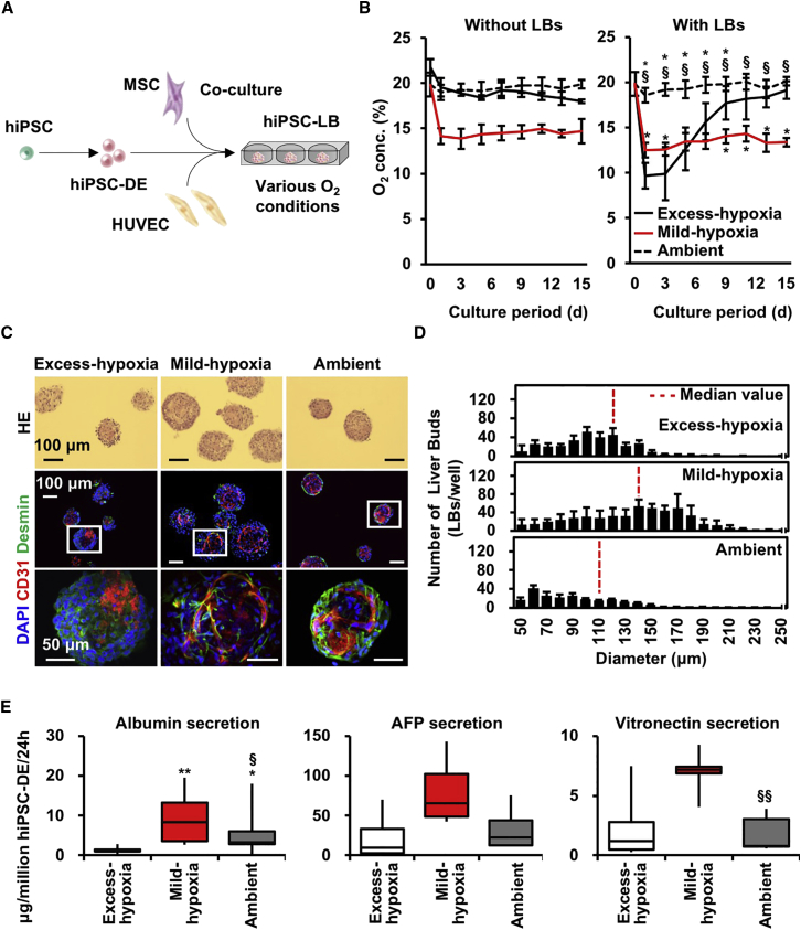
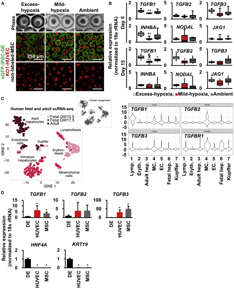
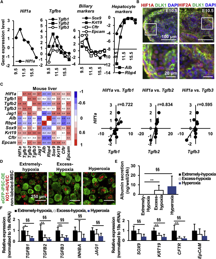
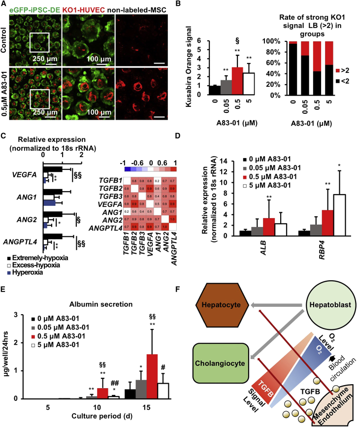
Timely controlled oxygen (O2) delivery is crucial for the developing liver. However, the influence of O2 on intercellular communication during hepatogenesis is unclear. Using a human induced pluripotent stem cell-derived liver bud (hiPSC-LB) model, we found hypoxia induced with an O2-permeable plate promoted hepatic differentiation accompanied by TGFB1 and TGFB3 suppression. Conversely, extensive hypoxia generated with an O2-non-permeable plate elevated TGFBs and cholangiocyte marker expression. Single-cell RNA sequencing revealed that TGFB1 and TGFB3 are primarily expressed in the human liver mesenchyme and endothelium similar to in the hiPSC-LBs. Stromal cell-specific RNA interferences indicated the importance of TGFB signaling for hepatocytic differentiation in hiPSC-LB. Consistently, during mouse liver development, the Hif1a-mediated developmental hypoxic response is positively correlated with TGFB1 expression. These data provide insights into the mechanism that hypoxia-stimulated signals in mesenchyme and endothelium, likely through TGFB1, promote hepatoblast differentiation prior to fetal circulation establishment.

Keywords: differentiation; hypoxia; iPSC; liver bud; organogenesis; organoid; oxygen.


Figures

Figure 1
Figure 1
Hypoxic Conditions Generated with an O2-Permeable Plate Promoted Hepatocyte Differentiation in Liver Buds (A) Schematic view of the present study. (B) Changes in O2 tension in culture medium without (left) or with (right) hiPSC-LBs for 15 days (mean ± SD; n = 8 or 9 independent experiments; p < 0.05 versus Excess-hypoxia; §p < 0.05 versus Mild-hypoxia). (C) H&E and immunofluorescence staining of hiPSC-LBs cultured for 15 days (CD31: red; Desmin: green; nuclei: blue [DAPI]). Scale bar, 100 μm (upper and middle side) or 50 μm (lower side). (D) Diameter distribution of hiPSC-LBs cultured for 15 days (mean ± SD; n = 9–11 independent experiments). (E) Boxplots of hiPSC-DE cell number normalized protein level in hiPSC-LB on day15. The error bars represent the maximum and minimum values; n = 8–18 independent experiments; p < 0.05 and ∗∗p < 0.01 versus Excess-hypoxia; §p < 0.05 and §§p < 0.01 versus Mild-hypoxia.
Figure 2
Figure 2
TGFB Signals from the Mesenchyme and Endothelium Are Candidate Regulators of O2-Dependent Hepatocyte Differentiation in Liver Buds (A) Phase-contrast and confocal images of hiPSC-LBs cultured for 1 (phase) or 5 (confocal) days (green: eGFP-iPSC-DE cells [AAVS1:EGFP]; red: KO1-HUVECs [MSCV-KO1]; no label: MSCs; scale bar, 250 μm). (B) Boxplots of TGFB family gene expression in hiPSC-LBs cultured for 5 and 15 days. The error bars represent the maximum and minimum values; n = 9 (day 5) and 10 (day 15) independent experiments; p < 0.05 and ∗∗p < 0.01 versus Excess-hypoxia; §p < 0.05 versus Ambient. (C) Left: tSNE of single-cell transcriptomes from human fetal (two donors, gestation weeks 10.5 and 17.5, 238 cells) and adult (three donors, age 21–65, 256 cells) liver samples (modified from Camp et al., 2017). Right: violin plots of single-cell transcriptomes, showing the distribution in the human liver. (D) Gene expression of each cell lineage in hiPSC-LB. hiPSC-LBs were cultured on Ambient group for 2 days. Then, dissociated eGFP-DE cells, KO-HUVECs, and unlabeled-MSCs from hiPSC-LBs were separated by FACS analysis using fluorescence labels. Gene expression of these cells was analyzed (mean ± SD; n = 8 independent experiments; p < 0.05 versus hiPSC-DE cells).
Figure 3
Figure 3
Hif1a Is Positively Correlated with Tgfbs and Biliary Markers and Negatively Correlated with Hepatocyte Markers in Mouse Liver Development (A) Microarray gene expression analysis of mouse fetal liver from E9.5 to 17.5. (B) Immunofluorescence staining for HIF1A (red), HIF2A (red), DLK1 (green), and nuclei (blue, DAPI) in E10.5 mouse liver. Scale bar, 100 μm (upper) or 20 μm (lower). (C) Correlation analysis of hypoxia- (Hif1a), hepatocyte- (Alb and Rbp4), and cholangiocyte- (others) associated markers in mouse livers from E9.5 to 8-week-old mice. (D) hiPSC-LBs cultured for 10 days (green: eGFP-iPSC-DE cells [AAVS1:EGFP]; red: KO1-HUVECs [MSCV-KO1]; no label: MSCs; scale bar, 250 μm). (E) ELISA on protein secretion in hiPSC-LBs cultured for 10 days (mean ± SD; n = 15 independent experiments; ∗∗p < 0.01 versus Excess-hypoxia, §§p < 0.01 versus Extreme-hypoxia). (F) Gene expression in hiPSC-LBs cultured for 10 days (mean ± SD; n = 12 independent experiments; p < 0.05 and ∗∗p < 0.01 versus Excess-hypoxia, §§p < 0.01 versus Extreme-hypoxia).
Figure 4
Figure 4
TGFB Signal Inhibition Promotes Hepatocyte Differentiation in Liver Buds (A) Confocal imaging of hiPSC-LBs cultured with various concentrations of A83-01 for 15 days in Excess-hypoxia group (green: eGFP-iPSC-DE cells [AAVS1:EGFP]; red: KO1-HUVECs [MSCV-KO1]; no label: MSCs; scale bar from left to right, 250, 100, and 100 μm). (B) Image analysis of HUVEC abundance in hiPSC-LBs cultured with various A83-01 concentrations for 15 days in Excess-hypoxia group. Fluorescence intensity of KO1 protein expression in HUVECs was evaluated as HUVEC abundance in hiPSC-LBs (left: mean ± SD; n = 9–17 independent experiments; ∗∗p < 0.01 versus 0 μM; §p < 0.05 versus 0.05 μM; right: a total of 1449–2985 LBs were measured). (C) Gene expression and correlation of hiPSC-LBs cultured for 10 days (mean ± SD; n = 12 independent experiments; p < 0.05 and ∗∗p < 0.01 versus Excess-hypoxia, §p < 0.05 and §§p < 0.01 versus Extreme-hypoxia). (D) Gene expression in hiPSC-LBs cultured with various A83-01 concentrations for 15 days in Excess-hypoxia group (mean ± SD; n = 6–14 independent experiments; p < 0.05 and ∗∗p < 0.01 versus 0 μM). (E) ELISA of protein secretion in hiPSC-LBs cultured with various A83-01 concentrations for 15 days in Excess-hypoxia group (mean ± SD; n = 9–17 independent experiments; p < 0.05 and ∗∗p < 0.01 versus 0 μM; §§p < 0.01 versus 0.05 μM, #p < 0.05 versus 0.5 μM; ##p < 0.05 versus 0.5 μM). (F) Putative mechanism of O2-dependent differentiation in human LBs through TGFB signaling.

References

    1. Anada T., Fukuda J., Sai Y., Suzuki O. An oxygen-permeable spheroid culture system for the prevention of central hypoxia and necrosis of spheroids. Biomaterials. 2012;33:8430–8441. - PubMed
    1. Anada T., Sato T., Kamoya T., Shiwaku Y., Tsuchiya K., Takano-Yamamoto T., Sasaki K., Suzuki O. Evaluation of bioactivity of octacalcium phosphate using osteoblastic cell aggregates on a spheroid culture device. Regen Ther. 2016;3:58–62. - PMC - PubMed
    1. Antoniou A., Raynaud P., Cordi S., Zong Y., Tronche F., Stanger B.Z., Jacquemin P., Pierreux C.E., Clotman F., Lemaigre F.P. Intrahepatic bile ducts develop according to a new mode of tubulogenesis regulated by the transcription factor SOX9. Gastroenterology. 2009;136:2325–2333. - PMC - PubMed
    1. Asai A., Aihara E., Watson C., Mourya R., Mizuochi T., Shivakumar P., Phelan K., Mayhew C., Helmrath M., Takebe T. Paracrine signals regulate human liver organoid maturation from induced pluripotent stem cells. Development. 2017;144:1056–1064. - PMC - PubMed
    1. Augustin H.G., Koh G.Y., Thurston G., Alitalo K. Control of vascular morphogenesis and homeostasis through the angiopoietin-Tie system. Nat. Rev. Mol. Cell Biol. 2009;10:165–177. - PubMed

Publication types

LinkOut - more resources