

Comparative investigation of porous nano-hydroxyapaptite/chitosan, nano-zirconia/chitosan and novel nano-calcium zirconate/chitosan composite scaffolds for their potential applications in bone regeneration
- PMID: 30033262
- PMCID: PMC6061966
- DOI: 10.1016/j.msec.2018.05.060
Comparative investigation of porous nano-hydroxyapaptite/chitosan, nano-zirconia/chitosan and novel nano-calcium zirconate/chitosan composite scaffolds for their potential applications in bone regeneration
Abstract
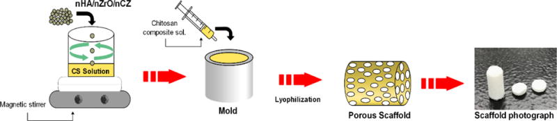
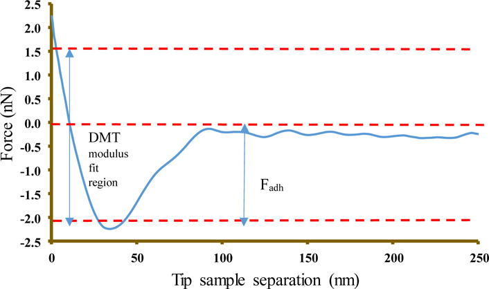
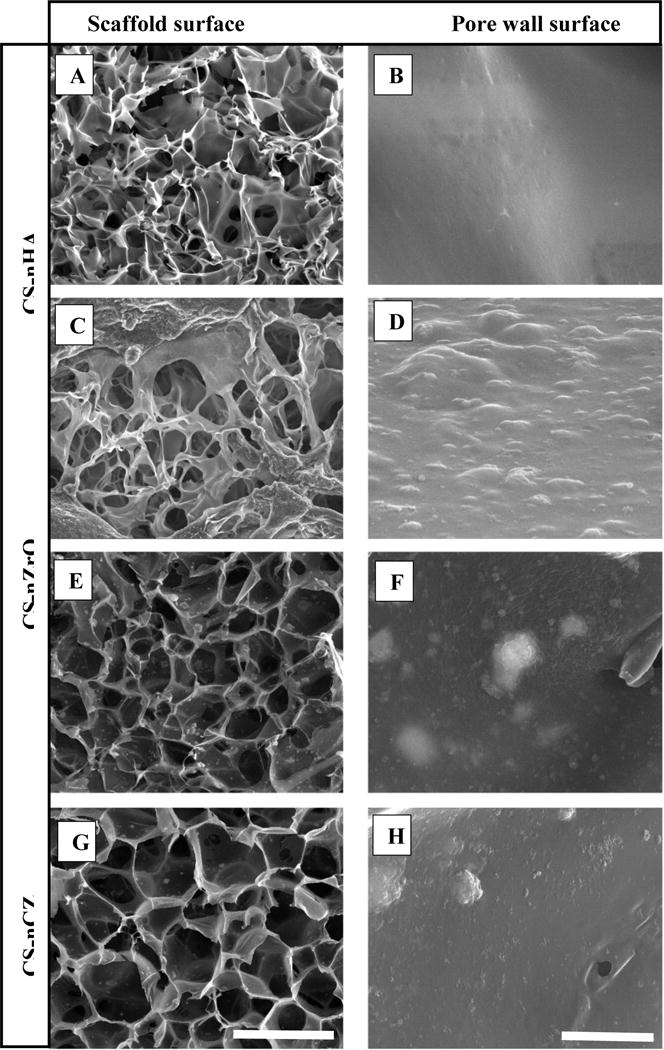
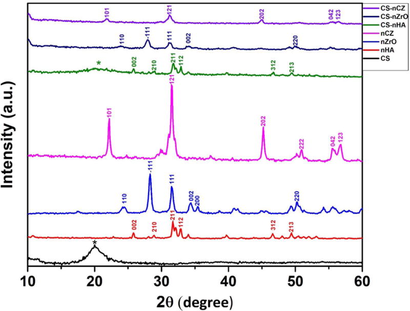
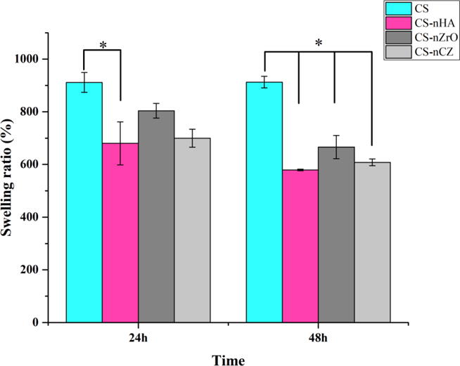
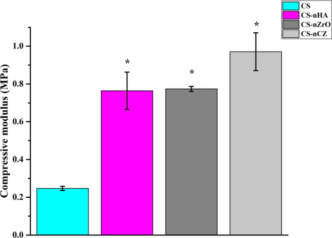
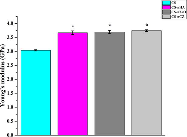
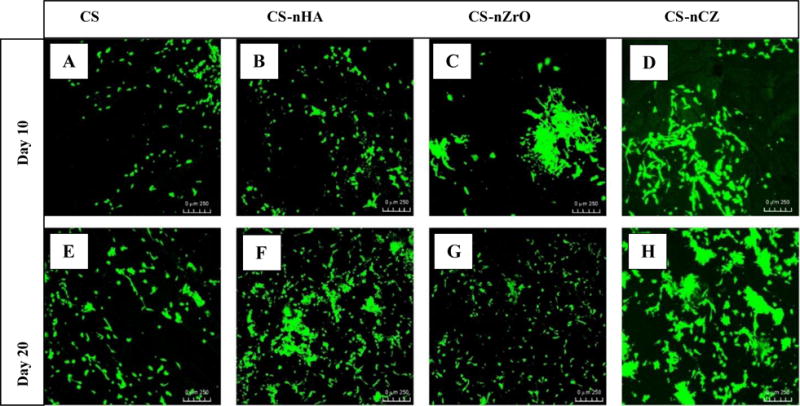
Zirconium (Zr) based bioceramic nanoparticles, as the filler material to chitosan (CS), for the development of composite scaffolds are less studied compared to hydroxyapatite nanoparticles. This is predominantly due to the biological similarity of nano-hydroxyapatite (nHA; Ca10(PO4)6(OH)2) with bone inorganic component. In this study, we compared the physical and biological properties of CS composite scaffolds hybridized with nHA, nano-zirconia (nZrO; ZrO2), and nano-calcium zirconate (nCZ; CaZrO3). For the first time in this study, the properties of CS-nCZ composite scaffolds have been reported. The porous composite scaffolds were developed using the freeze-drying technique. The compressive strength and modulus were in the range of 50-55 KPa and 0.75-0.95 MPa for composite scaffolds, significantly higher (p < 0.05), compared to CS alone scaffolds (28 KPa and 0.25 MPa) and were comparable among CS-nHA, CS-nZrO, and CS-nCZ scaffolds. Peak force quantitative nanomechanical mapping (PFQNM) using an atomic force microscope (AFM) showed that the Young's modulus of composite material was higher compared to only CS (p < 0.001), and the values were similar among the composite materials. One of the major issues in the use of Zr based bioceramic materials in bone tissue regeneration applications is their lower osteoblasts response. This study has shown that CS-nCZ supported higher proliferation of pre-osteoblasts compared to CS-nZrO and the spreading was more similar to that observed in CS-nHA scaffolds. Taken together, results show that the physical and biological properties, studied here, of CS composite with Zr based bio-ceramic was comparable with CS-nHA composite scaffolds and hence show the prospective of CS-nCZ for future bone tissue engineering applications.
Keywords: Atomic force microscopy; Cell proliferation; Chitosan; Composite; Mechanical properties; Nano-bioceramics.
Copyright © 2018 Elsevier B.V. All rights reserved.
Figures

References
-
- Stevens MM. Biomaterials for bone tissue engineering. Materials Today. 2008;11(5):18–25.
Publication types
MeSH terms
Substances
Grants and funding
LinkOut - more resources
Full Text Sources
Other Literature Sources
Miscellaneous
