. 2018 Aug 9;174(4):908-916.e12.
doi: 10.1016/j.cell.2018.05.058. Epub 2018 Jul 19.

Anti-CRISPR Phages Cooperate to Overcome CRISPR-Cas Immunity

Affiliations

Anti-CRISPR Phages Cooperate to Overcome CRISPR-Cas Immunity

Mariann Landsberger et al. Cell. .

Abstract

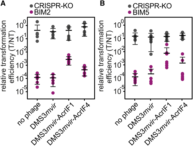
Some phages encode anti-CRISPR (acr) genes, which antagonize bacterial CRISPR-Cas immune systems by binding components of its machinery, but it is less clear how deployment of these acr genes impacts phage replication and epidemiology. Here, we demonstrate that bacteria with CRISPR-Cas resistance are still partially immune to Acr-encoding phage. As a consequence, Acr-phages often need to cooperate in order to overcome CRISPR resistance, with a first phage blocking the host CRISPR-Cas immune system to allow a second Acr-phage to successfully replicate. This cooperation leads to epidemiological tipping points in which the initial density of Acr-phage tips the balance from phage extinction to a phage epidemic. Furthermore, both higher levels of CRISPR-Cas immunity and weaker Acr activities shift the tipping points toward higher initial phage densities. Collectively, these data help elucidate how interactions between phage-encoded immune suppressors and the CRISPR systems they target shape bacteria-phage population dynamics.

Keywords: Allee effect; CRISPR-Cas; anti-CRISPR; bacteria; bifurcation; epidemiology; immunosuppression; partial resistance; phage; tipping points.


Figures

None
Graphical abstract
Figure 1
Figure 1
CRISPR-Cas Confers Partial Immunity to Acr-Phages (A) Efficiency of plaquing (EOP) of DMS3vir (white bars) on PA14 WT (completely sensitive to DMS3vir), BIM2 (one spacer targeting DMS3vir), and BIM5 (four spacers targeting DMS3vir); EOP of DMS3mvir (black bars), DMS3mvir-AcrIF1 (red bars), and DMS3mvir-AcrIF4 (blue bars) on PA14 WT (one spacer targeting DMS3mvir, DMS3mvir-AcrIF1, and DMS3mvir-AcrIF4), BIM2 (two spacers targeting DMS3mvir, DMS3mvir-AcrIF1, and DMS3mvir-AcrIF4), and BIM5 (five spacers targeting DMS3mvir, DMS3mvir-AcrIF1, and DMS3mvir-AcrIF4). Data correspond to the mean of six independent replicate experiments. Error bars represent 95% confidence intervals (CI). (B) Fitness of bacteria with CRISPR resistance (PA14 WT, BIM2, and BIM5) relative to a phage-sensitive CRISPR-KO strain in the absence of phage (green data points) or in the presence of phage DMS3mvir-AcrIF1 (red data points) or phage DMS3mvir-AcrIF4 (blue data points). Data correspond to the mean of six independent replicate experiments; error bars represent 95% CI. See also Figure S1.
Figure S1
Figure S1
Bacteria with CRISPR Resistance Are Partially Immune to JBD26 and JBD30, Respectively, Related to Figure 1 (A) Efficiency of plaquing (EOP) of JBD26, which encodes AcrIF4 (blue bars) and JBD30, which encodes AcrIF1 (red bars) on BIM4 (1 newly acquired spacer against both phages). Data correspond to the mean of 6 independent replicate experiments. Error bars represent 95% c.i. (B–E) Viral titers at 24 hours post-infection (hpi) with (B) JBD26 and (C) JBD30 on the CRISPR KO strain, (D) JBD26 and (E) JBD30 on BIM4. Gray circles indicate the phage titers (pfu/ml) at the start of the experiment (corresponding to the addition of 104, 105, 106, 107 or 108 pfus). Colored data points represent phage titers at 24 hpi. Note that the observed differences in amplification of JBD30 and JBD26 on CRISPR KO may be due to differences in their rates of lysogenisation. Each experiment was performed as 6 independent replicates, error bars represent 95% c.i.
Figure 2
Figure 2
The Initial MOI of Acr-Phage Determines the Epidemiological Outcome (A–L)Viral titers at 24 hr post-infection (hpi) with DMS3mvir (A, D, G, and J), DMS3mvir-AcrIF1 (B, E, H, and K), or DMS3mvir-AcrIF4 (C, F, I, and L) of PA14 CRISPR-KO (A–C), WT (D–F), BIM2 (G–I), or BIM5 (J–L). Gray circles indicate the phage titers at the start of the experiment (corresponding to the addition of 104, 105, 106, 107, or 108 pfus). Colored data points represent phage titers at 24 hpi; each data point represents an independent biological replicate (n = 6). The limit of detection is 200 pfu/ml. See also Figure S2.
Figure S2
Figure S2
Epidemiological Tipping Points Cannot Be Explained by Phage Evolution or Csy Complex Sequestration, Related to Figure 2 (A) Deep sequencing of protospacer sequences of phages DMS3mvir (black data points), DMS3mvir-AcrIF1 (red data points) or DMS3mvir-AcrIF4 (blue data points), either ancestral (A) or evolved on WT, BIM2 or BIM5 hosts (DMS3mvir only on WT). Protospacer 1 is targeted by WT, BIM2 and BIM5, protospacer 2 is targeted by BIM2 and BIM5, and protospacers 3, 4 and 5 are targeted by BIM5. Mean SNP frequency across the seed and PAM region (in total 10 nucleotides) of each protospacer is shown, error bars indicate the 95% c.i. (B) Density-dependent epidemiological tipping points are not due to phage evolution. Viral titers at 24 hpi of phage DMS3mvir (black data points), DMS3mvir-AcrIF1 (red data points) or DMS3mvir-AcrIF4 (blue data points) on bacteria PA14 WT, BIM2 or BIM5. Below each diagram is indicated which phage amounts (pfus) were added in the experiment. A indicates ancestral phage; E indicates evolved phage (isolated from the experiments depicted in Figure 2). (C) Viral titers at 24 hpi of BIM2 with 104, 105, 106, 107 or 108 pfus DMS3mvir-AcrIF1. (D) Viral titers at 24 hpi of BIM2 with 104, 105, 106, 107 or 108 pfus DMS3mvir-AcrIF1 in the presence of 108 pfus DMS3mvir. (E) Viral titers at 24 hpi of BIM2 with 104, 105, 106, 107 or 108 pfus DMS3mvir-AcrIF4. (F) Viral titers at 24 hpi of BIM2 with 104, 105, 106, 107 or 108 pfus DMS3mvir-AcrIF4 in the presence of 108 pfus DMS3mvir. Grey circles indicate the phage titers (pfu/ml) at the start of the experiment (corresponding to the addition of 104, 105, 106, 107 or 108 pfus). Colored points represent phage titers at 24 hpi; each data point represents an independent biological replicate (n = 6). The limit of detection is 200 pfu/ml.
Figure 3
Figure 3
Acr-Phage Amplification Is Density Dependent (A) Fold phage amplification at 24 hpi with 106 pfus DMS3mvir (black data points) or DMS3mvir-AcrIF1 (red data points) of PA14 CRISPR-KO (sensitive) or BIM2 under either high phage densities (HPD, 6 mL culture) or low phage densities (LPD, 600 mL culture). (B) Fold phage amplification at 24 hpi with 108 pfus DMS3mvir (black data points) or DMS3mvir-AcrIF4 (blue data points) of PA14 CRISPR-KO (sensitive) or BIM2 under either HPD or LPD. (C) Fold phage amplification at 24 hpi with 107 pfus DMS3mvir (black data points) or DMS3mvir-AcrIF1 (red data points) of PA14 CRISPR-KO (sensitive) or BIM5 under either HPD or LPD. (D) Fold phage amplification at 24 hpi with 108 pfus DMS3mvir (black data points) or DMS3mvir-AcrIF4 (blue data points) of PA14 CRISPR-KO (sensitive) or BIM5 under either HPD or LPD; each data point represents an independent biological replicate (n = 6). The limit of detection is 200 pfu/ml. See also Figure S3 and Table S1.
Figure S3
Figure S3
Surface Mutants Invade the Bacterial Population at High Acr-Phage Densities, Related to Figure 3 (A) Resistance phenotypes (C = CRISPR resistance, SM = surface resistance) that evolved at 48 hpi with phage DMS3vir-AcrIF1 at the indicated MOIs. All bacteria in the populations initially had CRISPR resistance (2 spacers targeting the phage). Phage amplification was observed at an MOI of 10−2 or higher. (B) Relative fitness of bacteria with CRISPR resistance (2 spacers targeting the phage) and surface resistance in the presence of phage DMS3vir-AcrIF1 at the indicated MOIs. (C) Resistance phenotypes (C = CRISPR resistance, SM = surface resistance) that evolved at 48 hpi with phage DMS3vir-AcrIF1 under either high phage densities (HPD, 6 mL culture) or low phage densities (LPD, 600 mL culture). All bacteria in the populations initially had CRISPR resistance (2 spacers targeting the phage). Data correspond to the mean of 6 independent replicate experiments. Error bars represent 95% c.i.
Figure 4
Figure 4
Epidemiological Tipping Points Can Result from Cooperation between Sequentially Infecting Acr-Phages (A) Infection model of the Acr-phage (see details of the model in STAR Methods). The parameter H(t) = αV(t) refers to the rate at which bacteria are infected by free phage particles. (B) Effect of initial Acr-phage inoculum density on the phage density at 24 hpi for different values of Acr efficacy (purple, ϕ = 0.65; magenta, ϕ = 0.55; green, ϕ = 0.35); other parameter values: B = 5, α = 0.001, ρ = 0.7, γ = 20. (C) Effect of initial Acr-phage inoculum density on the phage density at 24 hpi for different values of CRISPR efficacy (ρ = 0.5, 0.7, and 0.75; purple, magenta, green, respectively); other parameter values: B = 5, α = 0.001, ϕ = 0.6, γ = 20. (D) Effect of initial Acr-phage inoculum density on the phage density at 24 hpi for different values of the duration of the immunosuppressive state (γ = 0.1, 100, and 1,000; purple, magenta, green, respectively); other parameter values: B = 5, α = 0.001, ϕ = 0.65, ρ = 0.7. In all graphs, gray lines correspond to the initial amount of phage, and values below this line indicate a lack of phage amplification. See also Figure S4.
Figure S4
Figure S4
Partial Immunity Alone Cannot Explain the Observed Epidemiological Tipping Points, Related to Figure 4 Model predictions of the effect of initial Acr-phage inoculum density on the phage density at 24hpi for different values of Acr strength when no immunosuppressive state S is assumed in the model (ϕ = 0.67, 0.6 and 0.5; purple, magenta, green respectively); other parameter values: B = 5, α = 0.001, γ = 20, ρ = 0.7. Grey line corresponds to the initial amount of phage and values below this line indicate a lack of phage amplification.
Figure 5
Figure 5
Unsuccessful Infections by Acr-Phages Cause Hosts with CRISPR Resistance to Become Immunosuppressed (A) Relative transformation efficiencies (RTE) of CRISPR-KO (gray data points) or BIM2 (purple data points) pre-infected with 1.6 × 109 pfus of either DMS3mvir, DMS3mvir-AcrIF1, or DMS3mvir-AcrIF4 or not phage infected. Each data point represents an independent biological replicate (n = 6). (B) RTE of CRISPR-KO (gray data points) or BIM5 (purple data points) pre-infected as described for (A). Each data point represents an independent biological replicate (n = 7). In addition, we show the mean and 95% CI for each treatment. See also Figure S5 and Table S2.
Figure S5
Figure S5
Model Predictions of the Temporal Population Dynamics of Acr-Phage and Resistant and Immunosuppressed Hosts, Related to Figure 5 (A and B) Model predictions for the densities of resistant bacteria (black), immunosuppressed bacteria (orange) and phages (red) across time for two initial inoculum sizes: (A) V(0) = 5.104, (B) V(0) = 8.104. Other parameter values: R(0) = 106, B = 5, α = 0.001, γ = 20, ρ = 0.7. (C and D) Model predictions of the temporal population dynamics of Acr-phage and resistant and immunosuppressed hosts when the bacteria is initially composed of two different resistant hosts with different efficacy of resistance (ρ1 = 0.7 and ρ2 = 0.4) in equal frequency, with the densities of the two resistant bacteria (full and dashed black lines, respectively), immunosuppressed bacteria (orange) and phages (red) across time for two initial inoculum sizes: (C) V(0) = 5.104, (D) V(0) = 6.104. Other parameter values: B = 5, α = 0.001, γ = 20. (E) Effect of initial Acr-phage inoculum density on the phage density at 24 hpi for different values of Acr efficacy (ϕ = 0.42 (purple), ϕ = 0.35 (magenta) and ϕ = 0.2 (green)) when the bacteria is initially composed of two different resistant hosts with different efficacy of resistance (ρ1 = 0.7 and ρ2 = 0.4) in equal frequency. Other parameter values: B = 5, α = 0.001, γ = 20. (F) Viral titers at 24 hpi of a 50:50 mix of BIM2 and BIM5 with 104, 105, 106, 107 or 108 pfus DMS3mvir-AcrIF1. Grey circles indicate the phage titers (pfu/ml) at the start of the experiment. Colored points represent phage titers at 24 hpi; each data point represents an independent biological replicate (n = 6). The limit of detection is 200 pfu/ml.

Comment in

References

    1. Andersson A.F., Banfield J.F. Virus population dynamics and acquired virus resistance in natural microbial communities. Science. 2008;320:1047–1050. - PubMed
    1. Antia R., Regoes R.R., Koella J.C., Bergstrom C.T. The role of evolution in the emergence of infectious diseases. Nature. 2003;426:658–661. - PMC - PubMed
    1. Bankevich A., Nurk S., Antipov D., Gurevich A.A., Dvorkin M., Kulikov A.S., Lesin V.M., Nikolenko S.I., Pham S., Prjibelski A.D. SPAdes: a new genome assembly algorithm and its applications to single-cell sequencing. J. Comput. Biol. 2012;19:455–477. - PMC - PubMed
    1. Barrangou R., Fremaux C., Deveau H., Richards M., Boyaval P., Moineau S., Romero D.A., Horvath P. CRISPR provides acquired resistance against viruses in prokaryotes. Science. 2007;315:1709–1712. - PubMed
    1. Bondy-Denomy J., Garcia B., Strum S., Du M., Rollins M.F., Hidalgo-Reyes Y., Wiedenheft B., Maxwell K.L., Davidson A.R. Multiple mechanisms for CRISPR-Cas inhibition by anti-CRISPR proteins. Nature. 2015;526:136–139. - PMC - PubMed

Publication types

MeSH terms

LinkOut - more resources