. 2018 Jul 26;174(3):730-743.e22.
doi: 10.1016/j.cell.2018.06.019. Epub 2018 Jul 19.

A Complete Electron Microscopy Volume of the Brain of Adult Drosophila melanogaster

Affiliations

A Complete Electron Microscopy Volume of the Brain of Adult Drosophila melanogaster

Zhihao Zheng et al. Cell. .

Abstract

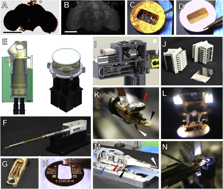
Drosophila melanogaster has a rich repertoire of innate and learned behaviors. Its 100,000-neuron brain is a large but tractable target for comprehensive neural circuit mapping. Only electron microscopy (EM) enables complete, unbiased mapping of synaptic connectivity; however, the fly brain is too large for conventional EM. We developed a custom high-throughput EM platform and imaged the entire brain of an adult female fly at synaptic resolution. To validate the dataset, we traced brain-spanning circuitry involving the mushroom body (MB), which has been extensively studied for its role in learning. All inputs to Kenyon cells (KCs), the intrinsic neurons of the MB, were mapped, revealing a previously unknown cell type, postsynaptic partners of KC dendrites, and unexpected clustering of olfactory projection neurons. These reconstructions show that this freely available EM volume supports mapping of brain-spanning circuits, which will significantly accelerate Drosophila neuroscience. VIDEO ABSTRACT.

Keywords: Drosophila melanogaster; connectomics; electron microscopy; image stitching; mushroom body; neural circuits; olfaction.


Figures

None
Graphical abstract
Figure 1
Figure 1
Target Volume and EM Acquisition Infrastructure (A) Oblique view of a surface model of the Drosophila brain (gray mesh) with specific neuropil compartments highlighted: AL (orange), MB (pink), and LH (green). (B) Schematic of olfactory pathway. Approximately 150 PNs, divided into ∼50 subtypes based on their anatomically defined glomeruli of origin in the AL, project to the MB calyx and LH (Grabe et al., 2016, Masse et al., 2009). Lateral horn is thought to mediate innate olfactory behaviors, whereas the MB is involved in learned behaviors (Keene and Waddell, 2007). In MB calyx, PN collaterals terminate in boutons and synapse on KCs (Yasuyama et al., 2002). Most PN types project to the MB calyx via the mALT, but several travel in secondary tracts (data not shown), and a few bypass calyx and project only to LH (Frank et al., 2015, Stocker et al., 1990, Tanaka et al., 2012). (C) Workflow for the generation of the whole-brain dataset. Blocks of brain tissue are incubated in heavy metals to label cell membranes, embedded in a resin polymer, and screened with X-ray tomography. Blocks were then serially sectioned with a diamond knife (for an introduction to serial sectioning techniques, see Harris et al., 2006). Groups of three serial sections are placed on metal slot grids for imaging in one of the two custom high-throughput TEM systems (TEMCA2 or ATPS). The imaged sections were assembled into an aligned volume with the custom software pipeline described here. Reconstruction and analyses of neural circuits in the volume were conducted with the CATMAID tracing environment (Saalfeld et al., 2009). D, dorsal; V, ventral; A, anterior; P, posterior; M, medial; L, lateral; LH, lateral horn; mALT, medial antennal lobe tract. See also Figures S1, S2, and S3 and Videos S1, S2, S3, and S4.
Figure S3
Figure S3
Sample Preparation and EM Acquisition Infrastructure, Related to Figure 1 (A) Drosophila brain following en bloc staining. (B) Frontal view of a 3D volumetric rendering of an X-ray tomogram from the embedded Drosophila brain. (C) Sample support film test showing a failed result with wrinkling of the support film on a 3 mm grid with 2 × 1 mm slot. (D) Sample support film test showing a successful result with no wrinkling or relaxation of the support film. (E) Left, schematic of TEMCA2 vacuum extension, scintillator, and camera array. Right, camera diagram showing the non-overlapping FOV of each camera on the scintillator. (F) An FEI CompuStage-compatible single-axis Fast Stage. (G) A Fast Stage grid holder. (H) A 3 mm grid with a 2 × 1 mm slot, custom-etched fiducial marks, 2D barcodes, and unique serial identifier. (I) Cassettes, magazine, and four-axis stage inside the ATPS vacuum. (J) ATPS cassettes and magazines. (K) Grid holder and end-effector of the ATPS grid positioning system. Arrows: prism and LED assembly (red); sample grid (black); lever of the grip assembly (white) which actuates grid release. (L) ATPS end-effector with LED lights for machine vision-guided pick-and-place. (M) Four-axis ATPS stage. Arrows: transverse precision piezo-driven axis (blue); pitch-axis pivot point (red); grid positioning system shuttle piezo motor (black); end effector and vision-system camera (white). (N) Rotational aligner integrated into the ATPS cassette shuttle orients the grids for imaging. Scale Bars: 250 μm in (A) and (B).
Figure S2
Figure S2
TEMCA2, Fast Stage, and ATPS for High-Throughput EM Imaging, Related to Figure 1 (A) A TEMCA2 equipped with a Fast Stage. Arrowheads: Fast Stage (black); elongated vacuum chamber (white); 2 × 2 camera array (red). (B) Fast Stage Schematic. Upper, driven mass (left) and exterior view (right). Lower, cutaway of Fast Stage showing the locations of dampers, bearings, and vacuum bellows. Arrows: rolling element damper locations (black arrows); rolling-element ‘tip’ bearing (white arrowhead); vacuum bellows (black arrowhead). (C) Plot of Fast Stage motion over time following an 8 μm move. Top trace, vibration perpendicular to Fast Stage motion axis. Bottom trace, vibration along the Fast Stage motion axis. (D) Schematic of Fast Stage stepping pattern. Left, small-step/big-step schematic. Numbers indicate camera identity within the array. Right, CompuStage and Fast Stage scanning axes. Red point is origin of scanning. (E) ATPS (white arrowhead) mounted to an accessory port on an FEI Tecnai Spirit BioTWIN TEM. (F) Schematic of the ATPS system diagraming motor positions and movement axes as well as vacuum and pneumatics subsystems.
Figure 2
Figure 2
Reconstructed Image Volume (A–F) Renderings of brain-spanning EM in the sectioning plane (x-y axes) at successive zoom levels. All panels rendered using the ELM viewer (STAR Methods), which averages several adjacent sections to improve contrast at low magnifications. Red dotted lines in left column indicate orthogonal (y-z axes) section plane through the whole-brain volume, rendered in right column. (G) Image signal-to-noise ratio (SNR) versus per-section acquisition rates for the current dataset (TEMCA2) and publicly available volume EM datasets acquired via comparable techniques: FIB-scanning EM (Takemura et al., 2015), SBEM (Briggman et al., 2011), ATUM-scanning EM (Kasthuri et al., 2015), ssTEM (Takemura et al., 2013), and TEMCA (Bock et al., 2011). Error bars indicate SD. (H–K) Serial thin sections succeeding the one in (F). Fine processes can be followed across serial sections and section-to-section image registration is accurate enough to provide a consistent FOV. Axis labels are the same as those used in Figure 1. Scale bars, 200 (A), 100 (B), 25 (C), 10 (D), 2 (E), and 0.4 μm (F and H–K). See also Figures S4, S5, and S6.
Figure S4
Figure S4
Comparison of SNR between EM Imaging Methods, Related to Figure 2 (A) Sample images from a variety of EM datasets acquired via different techniques. The data sources are as in Figure 2G. The top row shows images of side length 3 μm while the lower row shows 100 × 100 pixel subimages of each. Red squares indicate the areas of the subimages. (B) From left to right, a TEMCA2 image, the key-points detected in the image, convolution of the key-points illustrating dense and sparse feature regions (purple – low, yellow – high), the region of sparse features selected from the TEMCA2 image showing a resin filled area suitable for noise calculation. (C) The normalized SNR versus acquisition rates of a variety of EM techniques are shown for different SNR methods. Color code, points and data sources are as in Figure 2G. From left to right, the feature-based method is as described in (B); for the stacked voxels method, voxels are combined across a layer (SBEM not shown due to unclear alignments) and across 50 random images; for the reduced resolution method, voxels correspond to a larger physical size across 100 random images; for the scaled up resolution method, voxels correspond to a smaller physical size across 100 random images; for the Gaussian Blur method, voxels have been blurred with a Gaussian filter across 100 random images. Error bars indicate SD. (D) Cell membrane SNR method. Left, a representative image used to select two lines of pixels for quantifying signal (red line) and noise (green line), respectively. The pixels used for signal quantification were selected from cell membranes, and pixels used for noise quantification were selected from areas that contained only resin. Right, the grayscale values for signal (red) and noise (green) pixels selected in each region. (E) Normalized SNR versus acquisition rates as determined via the cell membrane method across five random images from each technique, each of which had 10 regions of background/noise and signal determined. Color code, points, and data sources are as in Figure 2G. Error bars indicate SD.
Figure S5
Figure S5
Re-imaging Synapses in MB Pedunculus, Montaging, 2D Intensity Correction, and Assessment of Volume Quality, Related to Figure 2 (A–C) Matching FOVs in section 3887 from the whole-brain volume (A) and re-imaged at higher resolution in (B-C). Resolutions in (A) and (B) and (C) are 4 nm/pixel and 0.5 nm/pixel, respectively. (D, G, and I) Whole-brain sections. (D–F) Registration of images acquired with high-dose and low-dose current beams. Debris present on a section (red rectangle) necessitated collection of a small subset of tiles at lower dose than the remainder of the mosaic. Red rectangle indicates the subregion displayed in (E). (E) A higher magnification image of the debris and border of the low-dose mosaic indicated in (D). (F) The boundary (arrowheads) in the joined high-dose and low-dose montage. (G–J) Mosaic of the same section prior to (G-H) and after (I-J) 2D intensity correction. Red squares in (G) and (I) indicate the subregions shown in (H) and (J), respectively. Intensity differences visible in (H) are greatly diminished in (J). (K) Most sections have few lost or degraded tiles. Red line, the running median of the total number of tiles per section for an 11-section window (five either side). For sections with lost tiles, only those with tile loss more than 5% below the median are shown. Triangles indicate complete loss of three consecutive sections. For sections containing degraded tiles, only those with 100 or more tiles contaminated by artifacts are shown. The dense data points and the fluctuations of running medians toward both ends of the series (sections before ∼1500 or after ∼5800) are due to tiles that contain extraneous tissue or resin outside the neuropil compartment. The tile count per section (running medians) across the series is proportional to the cross-sectional area of the brain normal to the cutting direction (z axis). (L) Volume alignment quality is sufficient for neural reconstruction. Alignment quality was assessed by analyzing the section-by-section displacements of individual Gaussian-smoothed skeletons of reconstructed neurons (STAR Methods). Large displacements are generally indicative of section misalignments. Data outside of a core region of the brain (sections < ∼500 and > ∼5500, shaded regions) are not informative since: 1) these regions mostly contain somata which typically have larger diameters than neurites, resulting in increased variability of tracing node placements, and 2) not enough tracing exists outside this range. The median and the 95% percentile of the displacements are 0.09 μm and the 0.57 μm, respectively. Scale Bars: 200 nm in (A)–(C), 100 μm in (D), (G), and (I), 25 μm in (E), 1 μm in (F), 2 μm in (H) and (J).
Figure S1
Figure S1
Neuronal Architecture of the MB Calyx, Related to Figure 1 (A) Micrograph of a microglomerulus in MB calyx. A canonical olfactory PN bouton (pink) is presynaptic to several fine KC dendrites, forming a synaptic complex referred to as a microglomerulus (Yasuyama et al., 2002). Arrows: presynaptic release sites. (B) Schematic of microglomerular inputs to KCs in MB calyx of Drosophila. The PN axons extend collaterals from the mALT into the calyx and provide bouton inputs to KCs. The Drosophila MB has ∼2,000 KCs on each side of the brain (Aso et al., 2014). Each KC projects a highly variable dendritic arbor into the calyx, which terminates in elaborations known as claws. Claws from many KCs wrap individual PN boutons to form each microglomerulus (Yasuyama et al., 2002), and each KC receives input from multiple PNs of diverse types across its claws (Caron et al., 2013, Gruntman and Turner, 2013). The complete composition of cell types that provide driving inputs via microglomeruli in the calyx is unknown. KCs have been shown to form presynaptic release sites in the calyx mostly outside of claws (Butcher et al., 2012, Christiansen et al., 2011), but the complete set of postsynaptic partners is unknown. Scale Bar: 1 μm in (A).
Figure S6
Figure S6
Reproducibility of Tracing, Related to Figure 2 (A and B) Three teams (indicated by colors), each comprising one tracer and one proofreader, reconstructed the same KC, with each team blinded to the others. (A) Morphologies are comparable across teams. Upper panel: asterisks indicate locations of a KC claw neurite postsynaptic to a PN input discovered by two out of three teams. Zoom-ins show the discrepancy of the fine claw neurites. Lower panel: cable length of missed branches for each of the three teams compared to an expert-generated gold-standard skeleton (STAR Methods). Consistent with Schneider-Mizell et al. (2016), the vast majority of our missed branches have a cable length of fewer than 5 μm. The reconstructed KC dendrite spanned serial sections 3451-4899. This range included one 3-section loss (3595-3597), and four single-section losses (3715, 4192, 4353, 4474), demonstrating the robustness of reproducible traceability to occasional data loss. (B) Synaptic annotations are comparable across teams. The KC (gray circle) receives input (arrows) from seven PN boutons (pale orange circles). The number of input synapses is indicated for each bouton, with the same team colors as in (A). All PN bouton inputs, except the rightmost one, were recapitulated by the three teams. Red asterisks mark the discrepant inputs caused by the missed branches in (A). Scale bars: ∼20 μm in (A), 250 nm in (A) inset.
Figure 3
Figure 3
Validation of Tracing by EM-LM Registration and NBLAST-Based Geometry Matching (A) An oblique cut plane through the EM volume, selected to reveal the projection from the AL to the MB calyx and LH via the mALT. The AL, mALT, MB calyx, and LH are false colored to show compartment boundaries. (B) The LM template brain is labeled with nc82 (magenta), a synapse-specific antibody commonly used to reveal neuropil compartment boundaries (Wagh et al., 2006). After alignment to the EM volume, the same cut plane reveals corresponding neuropil compartments in both (A) and here. Nc82 labeling is absent in the mALT, a largely synapse-free PN projection tract. (C) A subset of LM-imaged PNs labeled with random fluorophore combinations (Y. Aso, personal communication) using MultiColor FlpOut (Nern et al., 2015) were registered to the template brain, and the transformation defined in (B) was used to project the PNs into the coordinate space of the EM volume. The cut plane used in (A) reveals the PN dendrites in the AL, their axonal projections in the mALT, and their axonal arborizations in the MB calyx and LH. (D) Overlaid data from the EM dataset (A), the template brain (B), and the LM-imaged PNs (C) show good co-registration between the respective whole-brain image volumes. (E) An EM-reconstructed VM2 PN (black) is projected to a template brain (gray surface mesh) using the inverse of the transformation previously defined in (B) to align the LM template brain to the EM dataset. (F) An NBLAST search of the FlyCircuit database for matches to the EM-reconstructed VM2 PN (black) returned an LM-reconstructed VM2 PN (red) as the top hit. (G) An overlay of the EM- and LM-reconstructed VM2 PNs demonstrates high qualitative similarity. Axis labels as in Figure 1. Scale bars, ∼100 (A–D) and ∼50 μm (E–G).
Figure 4
Figure 4
Survey of Olfactory PNs Providing Driving Input to Microglomeruli in the MB Calyx Agrees with LM Data (A) EM-reconstructed uniglomerular olfactory PNs in the right hemisphere recapitulate known olfactory pathways (summarized in Figure 1B). (B) A frontal view of EM-reconstructed PNs (top) and glomerular surface models (bottom) in AL shows agreement with previous glomeruli reconstructions (Couto et al., 2005, Grabe et al., 2015). (C) A frontal-dorsal view of EM-reconstructed boutons for three groups of PNs in MB calyx reveals concentric organization, consistent with LM data (Tanaka et al., 2004). Bouton skeletons (top) were used to generate Gaussian-smoothed bouton volumes (bottom; STAR Methods) for each of the three groups. The PN groups are DM1, VA4, VC1, and VM2 (green); DL1 and VA6 (blue); and DA1, DC3, and VA1d (red). (D) Unsupervised clustering based on morphological similarity (NBLAST score) produces a dendrogram in which olfactory PNs are grouped by glomerular subtype. (E) Comparison of the number of reconstructed PNs per glomerulus from EM and LM data (Grabe et al., 2016). Colors in (A), (B), and (D) like in Couto et al. (2005). PNs receive input from olfactory receptor neurons (ORNs). The dendrite of each ORN innervates an antennal protuberance called a sensillum. Each PN is colored by the class of sensillum its input ORNs innervate. Error bars indicate SD. Axis and anatomical labels are the same as those used in Figure 1; lALT, lateral antennal lobe tract; LB, large basiconic; TB, thin basiconic; SB, small basiconic; T1, T2, T3, trichoid sensilla; PB, maxillary palp basiconic; AC, antennal coeloconic; AI, antennal intermediate. Scale bars, ∼10 μm (A–C). See also Table S1.
Figure 5
Figure 5
PN Arbors in MB Calyx Cluster More Tightly than Previously Seen with LM across Individuals (A) Comparison of EM- versus LM-reconstructed PNs. EM-reconstructed PNs are shown against a surface model of MB calyx (gray) in the left column. Calyx arbors for EM- and LM-reconstructed PNs are shown in the middle and right columns, respectively. Data for LM-reconstructed PNs (right column) are from the FlyCircuit database (Chiang et al., 2011), as registered to a common template brain (Costa et al., 2016; see also STAR Methods). (B) Pairwise distances between homotypic PN collaterals in the MB calyx. Each data point represents the distance between one pair of EM- (red) or LM-reconstructed (blue) PNs from the same subtype. Data points are bucketed according to PN subtype; subtypes are ordered on the x axis by how much more clustered EM-reconstructed PNs are than LM-reconstructed PNs (STAR Methods). (C) Histogram of all data points in (B). The mean of pairwise distances for all EM-reconstructed PN subtypes was significantly lower than that for all LM-reconstructed PN subtypes (3.40 ± 1.53 μm vs. 5.49 ± 2.73 μm, respectively; Student’s t test, p < 1.3 × 10−9). Axis labels as in Figure 1. Scale bars, ∼20 (A, left column) and ∼10 μm (A, middle and right columns). See also Figure S7.
Figure S7
Figure S7
Comparison of MB Calyx Collaterals Reveals Greater Similarity between EM-Reconstructed PNs than LM-Reconstructed PNs, Related to Figure 5 (A) Pairs of EM-reconstructed PNs are qualitatively more similar than pairs of LM-reconstructed PNs in the MB calyx. Subtypes are ordered by how much more clustered EM-reconstructed PNs are than LM-reconstructed PNs (STAR Methods). Middle column shows the pair of LM-reconstructed PNs with the median pairwise distance across all pairs. (B) Pairwise NBLAST scores between homotypic PN collaterals in the MB calyx. Each data point represents the NBLAST scores between one pair of EM- (red) or LM-reconstructed (blue) PNs. Data points are bucketed according to PN subtype; subtypes are ordered on the x axis by how much more similar EM-reconstructed PNs are than LM-reconstructed PNs (STAR Methods). (C) Histogram of all data points in (B). The mean of pairwise NBLAST scores for all EM-reconstructed PN subtypes was significantly higher than that for all LM-reconstructed PN subtypes (0.56 ± 0.18 versus 0.35 ± 0.21, respectively; Student’s t test, p < 2.2 × 10−12), indicating that EM-reconstructed PN subtypes are morphologically more similar to each other than LM-reconstructed PN subtypes. Scale bar: ∼10 μm in (A).
Figure 6
Figure 6
MB-CP2, a New Cell Type Providing Microglomerular Input to KC Claws (A) Reconstruction of the pair of MB-CP2 neurons with surface meshes of the whole brain (gray) and MB (green). (B–E) Synaptic connectivity between MB-CP2 and KCs in MB pedunculus and main calyx. Since many KC claws ensheath a given MB-CP2 bouton and KCs are traced only sparsely in this study, most postsynaptic profiles are untraced (STAR Methods). Arrowheads, presynaptic release sites. (B) A representative MB-CP2 neurite (orange) in MB pedunculus, postsynaptic to a KC axon (green). (C) A representative MB-CP2 neurite (orange) in left hemisphere MB pedunculus, with comparable synaptic arrangements to (B). KCs were not traced in the left hemisphere, so cell identity is putative. However, most bundled neurites parallel to the MB pedunculus long axis arise from KCs (Leitch and Laurent, 1996, Schürmann, 2016; data not shown). (D) A cross-section through a representative MB-CP2 bouton (orange) in MB main calyx at the center of a canonical microglomerulus. Several postsynaptic profiles arise from KC claws (green). (E) A representative MB-CP2 bouton in left hemisphere MB main calyx, with comparable synaptic arrangements to (D). KCs were not traced in left hemisphere, so cell identity is putative; however, most postsynaptic elements at MB calyx microglomeruli arise from KCs (Butcher et al., 2012; data not shown). (F) Summary schematic of MB-CP2 input and output brain regions with synapse counts discovered following partial reconstruction (STAR Methods). In six brain regions, MB-CP2 dendrites are purely postsynaptic. In four other regions, MB-CP2 neurites are both pre- and postsynaptic. Axis and anatomical labels as in Figure 1; Ped, MB pedunculus; dAC, dorsal accessory calyx; ATL, antler; SC, superior clamp; PLP, posterior lateral protocerebrum; SMP, superior medial protocerebrum; SIP, superior intermediate protocerebrum; SLP, superior lateral protocerebrum. Scale bars, 100 μm (A, dorsal view), 500 nm (B and C), and 2 μm (D and E). See also Video S5.
Figure 7
Figure 7
KC Presynaptic Release Sites in the MB Calyx Mostly Target a Small Subset of Available Partners (A–D) Morphological comparison of LM-imaged (left panels) and EM-reconstructed (right panels) neurons of the same class. LM data from Aso et al. (2014). Neurite densities are lower in the EM reconstructions, since these cells were traced to classification, not completion (STAR Methods). Spheres in the EM-reconstructions indicate the location of cell bodies. (A) αβc- (green), αβs- (yellow), and γ- (cyan) KCs. One representative EM-reconstructed KC from each class is shown (right panel); their morphologies and trajectories match those of the LM-imaged KCs of the same class. Small red square: location in MB calyx of a γ KC sub-arbor shown in the inset (large red square). Inset: representative location of a presynaptic release site (black arrowhead), on a twig arising from the KC backbone, outside the claw sub-arbor. A micrograph of this site is shown in (E). (B) APL, a wide-field inhibitory neuron that innervates the entire MB and sparsifies KC activity (Lin et al., 2014, Liu and Davis, 2009). (C) MB-CP1, a MB output neuron (MBON) with a dendritic arbor innervating the MB calyx and pedunculus (Tanaka et al., 2008). (D) MB-C1, a putative inhibitory interneuron that innervates the MB calyx and LH (Tanaka et al., 2008). Two MB-C1 neurons were found in the EM-based survey of KC postsynaptic targets, in contrast to the single neuron reported by Tanaka et al. (2008). Small red square: location in MB calyx of a γ KC sub-arbor shown in the inset (large red square). Inset: representative location of a presynaptic release site (black arrowhead), on a twig arising from the KC backbone, outside the claw sub-arbor. A micrograph of this site is shown in (F). For clarity, this KC is not shown in (D, right panel). (E and F) Micrographs at the synapse locations shown in (A) and (D) insets. Arrowheads, selected presynaptic release sites. (E) The γ KC in (A, inset) and two other γ KCs (light and dark purple) that are presynaptic to APL (green), MB-CP1 (red), and each other at the same synaptic cleft. The APL is also presynaptic to a PN (brown). (F) The γ KC from (D, inset) is presynaptic to MB-C1 (pink), APL (green), and several additional unidentified partners. The APL postsynaptic density is two sections away (not visible in this section plane). Scale bars, ∼25 (A–D) and 1 μm (E and F). See also Table S2.

Comment in

  • The whole fly brain in detail.
    Vogt N. Vogt N. Nat Methods. 2018 Sep;15(9):651. doi: 10.1038/s41592-018-0125-9. Nat Methods. 2018. PMID: 30171244 No abstract available.

Similar articles

  • Structured sampling of olfactory input by the fly mushroom body.
    Zheng Z, Li F, Fisher C, Ali IJ, Sharifi N, Calle-Schuler S, Hsu J, Masoodpanah N, Kmecova L, Kazimiers T, Perlman E, Nichols M, Li PH, Jain V, Bock DD. Zheng Z, et al. Curr Biol. 2022 Aug 8;32(15):3334-3349.e6. doi: 10.1016/j.cub.2022.06.031. Epub 2022 Jul 6. Curr Biol. 2022. PMID: 35797998 Free PMC article.
  • Presynaptic developmental plasticity allows robust sparse wiring of the Drosophila mushroom body.
    Elkahlah NA, Rogow JA, Ahmed M, Clowney EJ. Elkahlah NA, et al. Elife. 2020 Jan 8;9:e52278. doi: 10.7554/eLife.52278. Elife. 2020. PMID: 31913123 Free PMC article.
  • Complete Connectomic Reconstruction of Olfactory Projection Neurons in the Fly Brain.
    Bates AS, Schlegel P, Roberts RJV, Drummond N, Tamimi IFM, Turnbull R, Zhao X, Marin EC, Popovici PD, Dhawan S, Jamasb A, Javier A, Serratosa Capdevila L, Li F, Rubin GM, Waddell S, Bock DD, Costa M, Jefferis GSXE. Bates AS, et al. Curr Biol. 2020 Aug 17;30(16):3183-3199.e6. doi: 10.1016/j.cub.2020.06.042. Epub 2020 Jul 2. Curr Biol. 2020. PMID: 32619485 Free PMC article.
  • A connectome and analysis of the adult Drosophila central brain.
    Scheffer LK, Xu CS, Januszewski M, Lu Z, Takemura SY, Hayworth KJ, Huang GB, Shinomiya K, Maitlin-Shepard J, Berg S, Clements J, Hubbard PM, Katz WT, Umayam L, Zhao T, Ackerman D, Blakely T, Bogovic J, Dolafi T, Kainmueller D, Kawase T, Khairy KA, Leavitt L, Li PH, Lindsey L, Neubarth N, Olbris DJ, Otsuna H, Trautman ET, Ito M, Bates AS, Goldammer J, Wolff T, Svirskas R, Schlegel P, Neace E, Knecht CJ, Alvarado CX, Bailey DA, Ballinger S, Borycz JA, Canino BS, Cheatham N, Cook M, Dreher M, Duclos O, Eubanks B, Fairbanks K, Finley S, Forknall N, Francis A, Hopkins GP, Joyce EM, Kim S, Kirk NA, Kovalyak J, Lauchie SA, Lohff A, Maldonado C, Manley EA, McLin S, Mooney C, Ndama M, Ogundeyi O, Okeoma N, Ordish C, Padilla N, Patrick CM, Paterson T, Phillips EE, Phillips EM, Rampally N, Ribeiro C, Robertson MK, Rymer JT, Ryan SM, Sammons M, Scott AK, Scott AL, Shinomiya A, Smith C, Smith K, Smith NL, Sobeski MA, Suleiman A, Swift J, Takemura S, Talebi I, Tarnogorska D, Tenshaw E, Tokhi T, Walsh JJ, Yang T, Horne JA, Li F, Parekh R, Rivlin PK, Jayaraman V, Costa M, Jefferis GS, Ito K, Saalfeld S, George R, Meinertzhagen IA, Rubin GM, Hess HF, Jain V, Plaza SM. Scheffer LK, et al. Elife. 2020 Sep 7;9:e57443. doi: 10.7554/eLife.57443. Elife. 2020. PMID: 32880371 Free PMC article.
  • Of what use is connectomics? A personal perspective on the Drosophila connectome.
    Meinertzhagen IA. Meinertzhagen IA. J Exp Biol. 2018 May 20;221(Pt 10):jeb164954. doi: 10.1242/jeb.164954. J Exp Biol. 2018. PMID: 29784759 Review.

Cited by

References

    1. Arganda-Carreras I., Kaynig V., Rueden C., Eliceiri K.W., Schindelin J., Cardona A., Sebastian Seung H. Trainable Weka segmentation: a machine learning tool for microscopy pixel classification. Bioinformatics. 2017;33:2424–2426. - PubMed
    1. Ashburner M., Golic K.G., Hawley R.S. Cold Spring Harbor Laboratory Press; 2005. Drosophila: A Laboratory Handbook.
    1. Aso Y., Hattori D., Yu Y., Johnston R.M., Iyer N.A., Ngo T.T., Dionne H., Abbott L.F., Axel R., Tanimoto H., Rubin G.M. The neuronal architecture of the mushroom body provides a logic for associative learning. eLife. 2014;3:e04577. - PMC - PubMed
    1. Bargmann C.I., Marder E. From the connectome to brain function. Nat. Methods. 2013;10:483–490. - PubMed
    1. Bay H., Ess A., Tuytelaars T., Van Gool L. Speeded-up robust features (SURF) Comput. Vis. Image Underst. 2008;110:346–359.

Publication types