

The Human Vomeronasal (Jacobson's) Organ: A Short Review of Current Conceptions, With an English Translation of Potiquet's Original Text
- PMID: 30034965
- PMCID: PMC6050168
- DOI: 10.7759/cureus.2643
The Human Vomeronasal (Jacobson's) Organ: A Short Review of Current Conceptions, With an English Translation of Potiquet's Original Text
Abstract
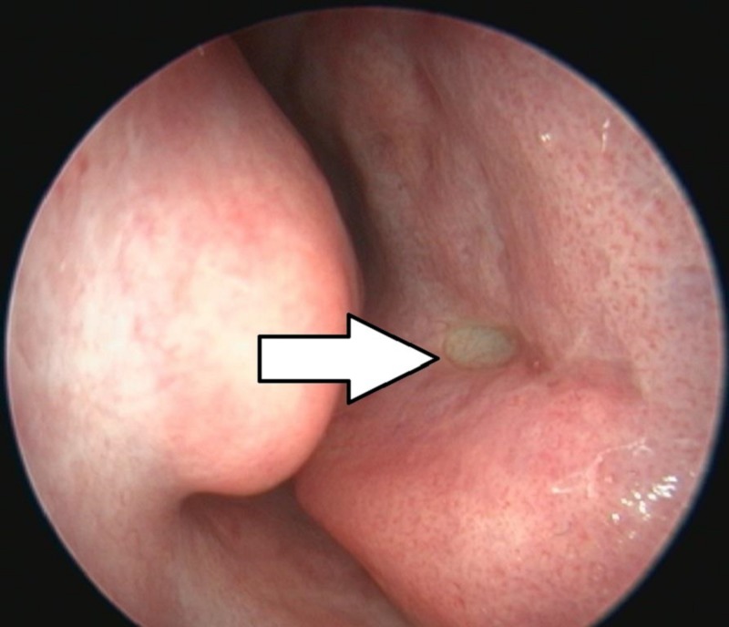
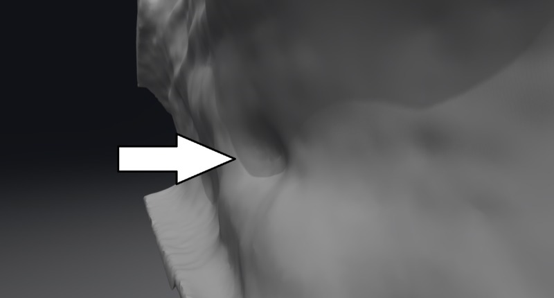
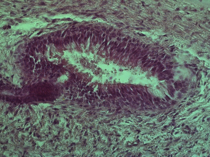
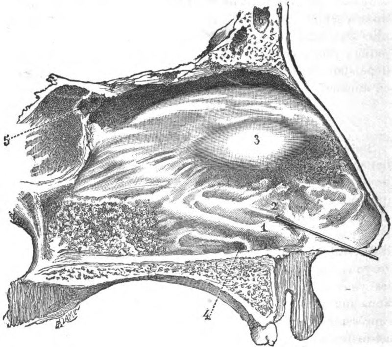
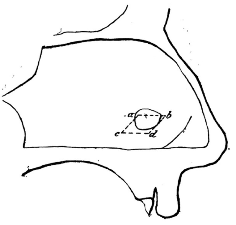
The vomeronasal organ (VNO) is a structure located in the anteroinferior portion of the nasal septum and is part of the accessory olfactory system. The VNO, together with its associated structures, has been shown to play a role in the formation of social and sexual behavior in animals, thanks to its pheromone receptor cells and the stimulating effect on the secretion of gonadotropin-releasing hormone. The VNO was first described as a structure by the Dutch botanist and anatomist Frederik Ruysch in 1703 while dissecting a young male cadaver. This finding, however, is widely contradicted due to no elaborate descriptions being made by the Ruysch. The description of the VNO is more widely attributed to the Danish surgeon Ludwig Jacobson, with whom the VNO has been synonymized, as in 1803 he described the structure in a variety of mammals. Whilst Jacobson extensively studied prior reports of the VNO, he publicly denied its existence in humans. Following these discoveries and some contradictory statements in 1891, M. Potiquet published one of the more influential reviews on the topic. To this day, despite the first report of the organ's existence being made in a human and many articles stating its presence and supporting its function, the presence of a VNO in humans is still widely debated upon.
Keywords: history; jacobson's organ; m. potiquet; vomeronasal organ.
Conflict of interest statement
The authors have declared that no competing interests exist.
Figures
References
-
- The human vomeronasal organ. V. An interpretation of its discovery by Ruysch, Jacobson, or Kölliker, with an English translation of Kölliker (1877) Bhatnagar KP, Smith TD. Anat Rec Part B New Anat. 2003;270:4–15. - PubMed
-
- Über die Entwicklung des Riechlappens und des Riechganglions und über diejenige des verlängerten Markes [Article in German] His W. Verh Anat Ges. 1889;3:63–66.
-
- The vomeronasal organ of the male ferret. Weiler E, Apfelbach R, Farbman AI. Chem Senses. 1999;24:127–136. - PubMed
-
- Puberty acceleration in mice. II. Evidence that the vomeronasal organ is a receptor for the primer pheromone in male mouse urine. Kaneko N, Debski EA, Wilson MC, Whitten WK. Biol Reprod. 1980;22:873–878. - PubMed
Publication types
LinkOut - more resources
Full Text Sources
Other Literature Sources