Review
. 2018 Jun 21:2018:3606970.
doi: 10.1155/2018/3606970. eCollection 2018.

Primary CNS Lymphomas: Challenges in Diagnosis and Monitoring

Affiliations
Review

Primary CNS Lymphomas: Challenges in Diagnosis and Monitoring

C Chiavazza et al. Biomed Res Int. .

Abstract

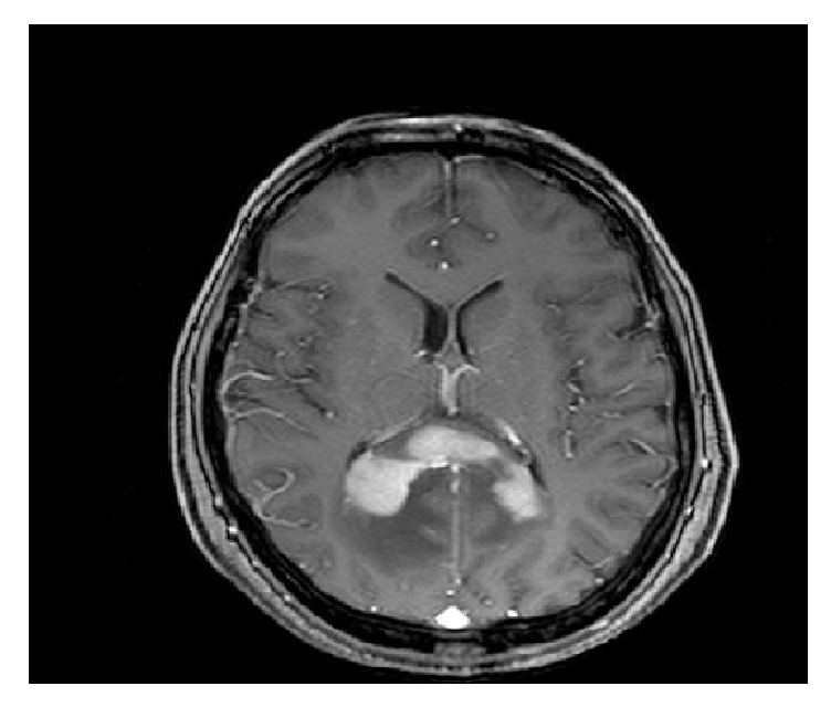
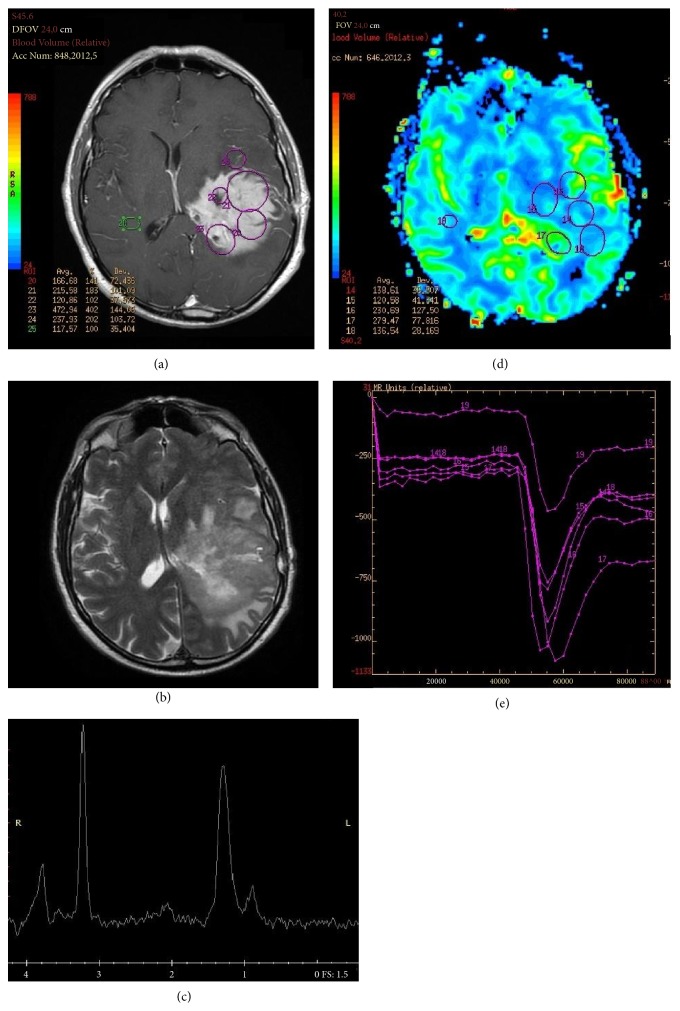
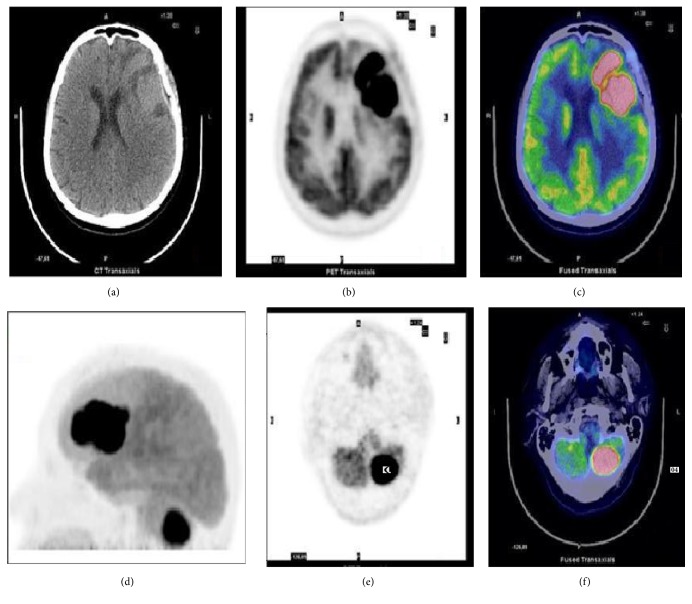
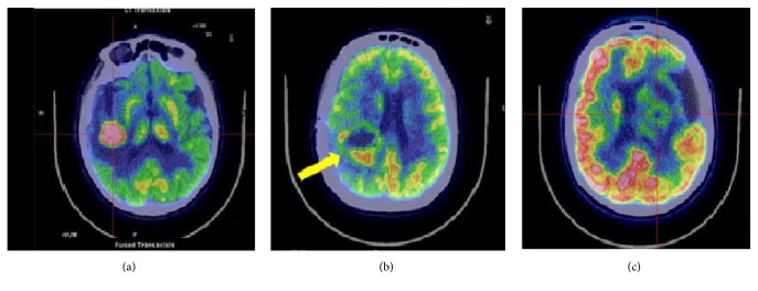
Primary Central Nervous System Lymphoma (PCNSL) is a rare neoplasm that can involve brain, eye, leptomeninges, and rarely spinal cord. PCNSL lesions most typically enhance homogeneously on T1-weighted magnetic resonance imaging (MRI) and appear T2-hypointense, but high variability in MRI features is commonly encountered. Neurological symptoms and MRI findings may mimic high grade gliomas (HGGs), tumefactive demyelinating lesions (TDLs), or infectious and granulomatous diseases. Advanced MRI techniques (MR diffusion, spectroscopy, and perfusion) and metabolic imaging, such as Fluorodeoxyglucose Positron Emission Tomography (FDG-PET) or amino acid PET (usually employing methionine), may be useful in distinguishing these different entities and monitoring the disease course. Moreover, emerging data suggest a role for cerebrospinal fluid (CSF) markers in predicting prognosis and response to treatments. In this review, we will address the challenges in PCNSL diagnosis, assessment of response to treatments, and evaluation of potential neurotoxicity related to chemotherapy and radiotherapy.


Figures

Figure 1
Figure 1
Contrast-enhanced axial T1-weighted MRI showing a PCNSL located in corpus callosum.
Figure 2
Figure 2
(a) Left temporoparietal PCNSL characterized by a homogeneous enhancing lesion on T1-weighted and (b) relatively low and inhomogeneous T2 signal on T2-weighted MRI. (c) Increased lipid peak on MRI spectroscopy and (d-e) increase of the regional cerebral blood volume (rCBV) when compared to the contralateral hemisphere.
Figure 3
Figure 3
18F-FDG brain imaging in a 66-year-old woman with PCNSL. Axial CT (a), PET (b), and PET/CT fusion image (c) showing the FDG-avid lesion involving the left frontal lobe (SUVmax 42). (d) Maximum imaging projection (MIP), axial CT (e), and PET/CT fusion image (f) showing another lesion on the left cerebellar hemisphere.
Figure 4
Figure 4
Axial 18F-FDG brain imaging. PCNSL (a), glioblastoma multiforme (b), and abscess (c).

References

    1. Villano J. L., Koshy M., Shaikh H., Dolecek T. A., McCarthy B. J. Age, gender, and racial differences in incidence and survival in primary CNS lymphoma. British Journal of Cancer. 2011;105(9):1414–1418. doi: 10.1038/bjc.2011.357. - DOI - PMC - PubMed
    1. Carnevale J., Rubenstein J. L. The challenge of primary central nervous system lymphoma. Hematology/Oncology Clinics of North America. 2016;30(6):1293–1316. doi: 10.1016/j.hoc.2016.07.013. - DOI - PMC - PubMed
    1. Montserrat E. Primary CNS lymphoma: in search of the evidence. The Lancet Haematology. 2015;2(6):e227–e228. doi: 10.1016/S2352-3026(15)00091-5. - DOI - PubMed
    1. Paydas S. Primary central nervous system lymphoma: essential points in diagnosis and management. Medical Oncology. 2017;34(4, article 61) doi: 10.1007/s12032-017-0920-7. - DOI - PubMed
    1. Lai R., Rosenblum M. K., DeAngelis L. M. Primary CNS lymphoma: a whole-brain disease? Neurology. 2002;59(10):1557–1562. doi: 10.1212/01.WNL.0000034256.20173.EA. - DOI - PubMed

MeSH terms

Substances