. 2018 Jun;1(3):e201800088.
doi: 10.26508/lsa.201800088. Epub 2018 Jun 18.

Silica-based solid-phase extraction of cross-linked nucleic acid-bound proteins

Affiliations

Silica-based solid-phase extraction of cross-linked nucleic acid-bound proteins

Claudio Asencio et al. Life Sci Alliance. 2018 Jun.

Abstract

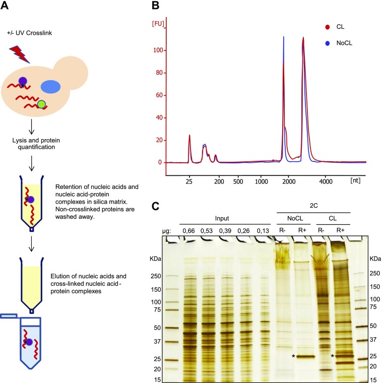
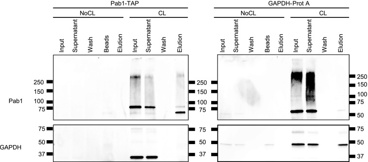
Proteins interact with nucleic acids to regulate cellular functions. The study of these regulatory interactions is often hampered by the limited efficiency of current protocols to isolate the relevant nucleic acid-protein complexes. In this report, we describe a rapid and simple procedure to highly enrich cross-linked nucleic acid-bound proteins, referred to as "2C" for "complex capture." This method is based on the observation that silica matrix-based columns used for nucleic acid purification also effectively retain UV cross-linked nucleic acid-protein complexes. As a proof of principle, 2C was used to isolate RNA-bound proteins from yeast and mammalian Huh7 cells. The 2C method makes RNA labelling redundant, and specific RNA-protein interactions can be observed and validated by Western blotting. RNA-protein complexes isolated by 2C can subsequently be immunoprecipitated, showing that 2C is in principle compatible with sensitive downstream applications. We suggest that 2C can dramatically simplify the study of nucleic acid-protein interactions and benefit researchers in the fields of DNA and RNA biology.


Conflict of interest statement

Conflict of Interest Statement The authors declare that they have no conflict of interest.

Figures

Figure 1.
Figure 1.. Isolation of RBPs from S. cerevisiae by 2C.
(A) Schematic representation of the 2C method. (B) Analysis of RNA integrity of cross-linked (red) and non–cross-linked (blue) yeast samples after irradiation of the cells with 3 J/cm2 of UV light at 254 nm and 2C extraction. (C) Visualization of yeast RBPs. 2C eluate samples equivalent to 12.5 μg of RNA were treated or not with RNase I, boiled for 5 min in loading buffer, and separated through a 4%–15% gradient SDS–PAGE subjected to silver staining. We noticed that the background contamination seen in the “NoCL” samples can be virtually eliminated by preincubation of the lysates at 70°C for 5 min. R−, non–RNase-treated samples; R+, RNase I–treated samples; *RNase I protein.
Figure 2.
Figure 2.. Validation of specific RNA–protein interactions from yeast and mammalian cells isolated by 2C.
(A) Evaluation of 2C performance examining known yeast RBPs by Western blotting. 2C eluates equivalent to 12.5 μg of RNA were treated or not with RNase I, boiled in loading buffer, and separated through SDS–PAGE. Specific RBPs were visualized with antibodies against Pab1 and GAPDH. The DNA-binding histone H3 was probed as a negative control. (B) Validation of additional non-canonical yeast RBPs by Western blot after 2C. The samples were treated as in (A), and hexokinase, triose phosphate isomerase, and tubulin, as a negative control, were analyzed with specific primary antibodies. Note that S. cerevisiae hexokinase B is known to aggregate under denaturing conditions in vitro into amyloid-like fibrils (Ramshini et al, 2011). The denaturing conditions during 2C capture, thus, may have promoted the formation of hexokinase aggregates resistant to SDS–PAGE separation. (C) Analysis of mammalian 2C eluates by Western blot. The samples were treated as above (A), and the proteins hnRNPC1/C2, GAPDH, FASTKD4, and histone H3 were detected with specific primary antibodies. R−, non–RNase-treated samples; R+, RNase I–treated samples.
Figure 3.
Figure 3.. Affinity purification of Pab1–RNA and GAPDH–RNA complexes after 2C.
RNPs of PAB1-TAP and GAPDH-ProtA strains were isolated by the 2C method. Eluates from 2C were subsequently affinity purified via the tags. Fractions from input, supernatant, washes, and elution were collected, subjected to Western blot analysis, and probed with antibodies against Pab1 and GAPDH.
Figure 4.
Figure 4.. Proposed applications of 2C in RNA biology.
For details, see the Discussion section. Also note that we envisage corresponding applications for DNA-binding proteins.

References

    1. Amberg D, Burke D, Strathern J (2005) Methods in Yeast Genetics: A Cold Spring Harbor Laboratory Course Manual, 2005 Edition. Cold Spring, NY: Cold Spring Harbor Laboratory Press
    1. Avison M. (2008) Measuring Gene Expression. London, UK: Taylor & Francis
    1. Baltz AG, Munschauer M, Schwanhausser B, Vasile A, Murakawa Y, Schueler M, Youngs N, Penfold-Brown D, Drew K, Milek M, et al. (2012) The mRNA-bound proteome and its global occupancy profile on protein-coding transcripts. Mol Cell 46: 674–690. 10.1016/j.molcel.2012.05.021 - DOI - PubMed
    1. Battaglia S, Lidschreiber M, Baejen C, Torkler P, Vos SM, Cramer P (2017) RNA-dependent chromatin association of transcription elongation factors and Pol II CTD kinases. Elife 6: 1–26. 10.7554/elife.25637 - DOI - PMC - PubMed
    1. Beckmann BM, Horos R, Fischer B, Castello A, Eichelbaum K, Alleaume AM, Schwarzl T, Curk T, Foehr S, Huber W, et al. (2015) The RNA-binding proteomes from yeast to man harbour conserved enigmRBPs. Nat Commun 6: 10127 10.1038/ncomms10127 - DOI - PMC - PubMed