. 2018 Jul 21;8(7):554.
doi: 10.3390/nano8070554.

Neurogenic Differentiation of Human Dental Pulp Stem Cells on Graphene-Polycaprolactone Hybrid Nanofibers

Affiliations

Neurogenic Differentiation of Human Dental Pulp Stem Cells on Graphene-Polycaprolactone Hybrid Nanofibers

Hoon Seonwoo et al. Nanomaterials (Basel). .

Abstract

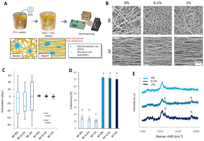
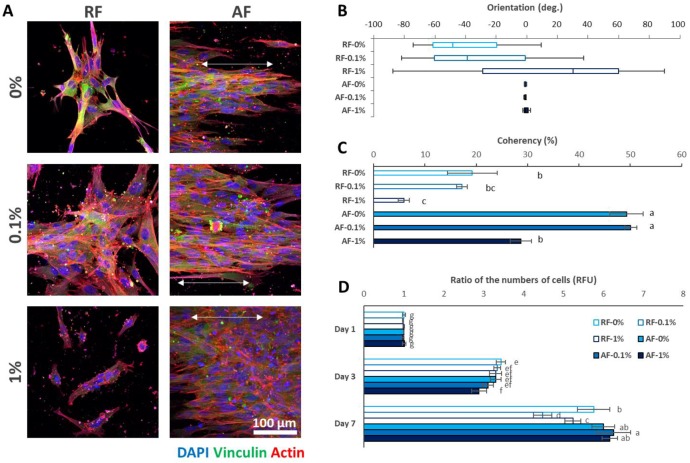
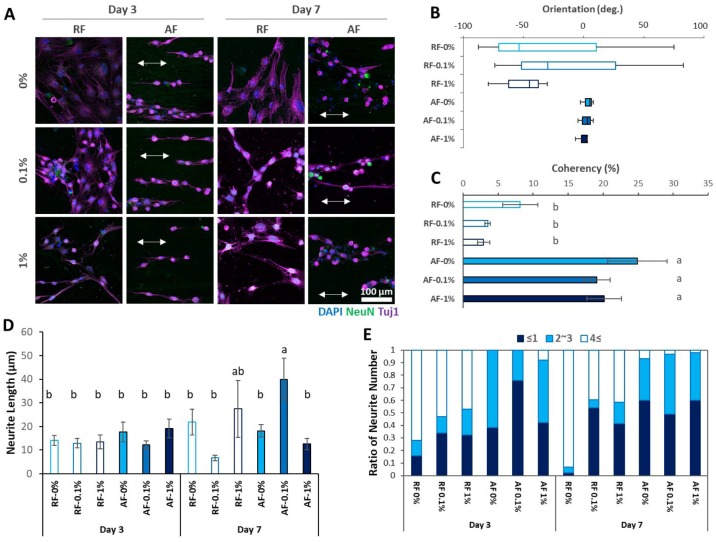
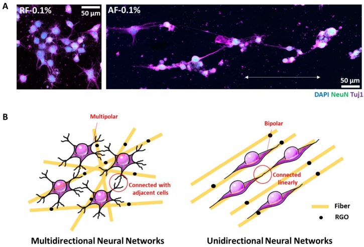
Stem cells derived from dental tissues-dental stem cells-are favored due to their easy acquisition. Among them, dental pulp stem cells (DPSCs) extracted from the dental pulp have many advantages, such as high proliferation and a highly purified population. Although their ability for neurogenic differentiation has been highlighted and neurogenic differentiation using electrospun nanofibers (NFs) has been performed, graphene-incorporated NFs have never been applied for DPSC neurogenic differentiation. Here, reduced graphene oxide (RGO)-polycaprolactone (PCL) hybrid electrospun NFs were developed and applied for enhanced neurogenesis of DPSCs. First, RGO-PCL NFs were fabricated by electrospinning with incorporation of RGO and alignments, and their chemical and morphological characteristics were evaluated. Furthermore, in vitro NF properties, such as influence on the cellular alignments and cell viability of DPSCs, were also analyzed. The influences of NFs on DPSCs neurogenesis were also analyzed. The results confirmed that an appropriate concentration of RGO promoted better DPSC neurogenesis. Furthermore, the use of random NFs facilitated contiguous junctions of differentiated cells, whereas the use of aligned NFs facilitated an aligned junction of differentiated cells along the direction of NF alignments. Our findings showed that RGO-PCL NFs can be a useful tool for DPSC neurogenesis, which will help regeneration in neurodegenerative and neurodefective diseases.

Keywords: alignments; dental pulp stem cells; nanofiber; neurogenesis; reduced graphene oxide.


Conflict of interest statement

The authors declare no conflict of interest.

Figures

Figure 1
Figure 1
Characteristics of the reduced graphene oxide (RGO)–polycaprolactione (PCL) hybrid electrospun nanofibers (NFs) (RGO-PCL NFs). (A) Study strategy. RGO was incorporated into PCL solution and sonicated for even distribution. Then, the solution was electrospun to achieve random nanofibers (RFs) and aligned nanofibers (AFs). The fabricated nanofibers were used for the neurogenic differentiation of dental pulp stem cells (DPSCs). (BD) Morphological analysis. (B) Representative images of electrospun RGO-PCL NFs. (C) Orientation of the RGO-PCL NFs. The orientation of the RFs are distributed broadly, whereas those of the AFs show narrow distribution. Each of the 25 images were used for the analysis. (D) Coherency of the RGO-PCL NFs. The coherencies of AFs were significantly higher than those of RFs. Each of 25 images was used for the analysis. Error bars represent the standard deviation. Same alphabets represent non-significance (p < 0.05). (E) Raman spectroscopy results. The 0.1% and 1% RGO-incorporated NFs exhibited D (~1450 cm−1) and G peaks (~1600 cm−1), the characteristic peaks of RGO. (C) XRD results. The 0.1% and 1%-incorporated NFs exhibited the characteristic peaks of RGO at approximately 23.5°.
Figure 2
Figure 2
Effects of the RGO-PCL NFs on DPSC behavior. (AC) Analyses of cellular morphologies. (A) Immunocytochemistry (ICC) results 2 days after seeding. Corresponding with the NF alignments, the DPSCs on the RFs were randomly aligned, whereas those on the AFs were aligned well. In particular, vinculin, an indicator of focal adhesion, did not stain the cells on the 1% RGO-PCL NFs, which indicated that excessive RGO incorporation may be harmful to initial cell adhesion. (B) DPSC alignments. Corresponding to the RGO-PCL NFs, the cells on the RFs showed broad orientation, whereas those on the AFs showed narrow orientation. (C) Coherency of the DPSC alignments. The alignments of the DPSCs on AFs were significantly higher than those of cells on the RFs, except for AF-1%. Furthermore, the cells on NFs with high RGO incorporation (1%) showed significantly decreased coherency. Error bars indicate the standard deviation. Same alphabets mean a non-significant difference between samples (p < 0.05). (D) Ratio of the numbers of DPSCs on the RGO-PCL NFs. On day 3, cells on AF-1% showed significantly decreased viability. On day 7, cells on AF-0.1% showed significantly higher viability, whereas those on RF-0.1% and RF-1% showed significantly lower viability. Error bars indicate the standard deviation. Same alphabets mean a non-significant difference between samples (p < 0.05).
Figure 3
Figure 3
Neurogenic differentiation of DPSCs using RGO-PCL NFs. (A) ICC results. The RGO-incorporated NFs resulted in faster morphological transformation and early expression of Tuj-1 and NeuN. The DPSCs on the RFs seemed randomly aligned, whereas those on the AFs seemed well-aligned. (B) Orientation of the alignments of the differentiated cells. Corresponding to the alignments of the RGO-PCL NFs, the cells on the RFs showed broad orientation, whereas those on the AFs showed narrow orientation. (C) Coherency of the alignments of the differentiated cells. The alignments of DPSCs on the AFs were significantly higher than those of the DPSCs on the RFs, except for AF-1%. Furthermore, the cells with high RGO incorporation (1%) showed significantly decreased coherency. Error bars indicate the standard deviation. Same alphabets mean a non-significant difference between samples (p < 0.05). (D) Neurite length of the differentiated cells. The AF-0.1% group showed significantly increased neurite length compared with other groups. Error bars indicate the standard deviation. Same alphabets mean a non-significant difference between samples (p < 0.05). (E) Distribution of neurite numbers on each group. RF groups and AF groups showed apparent changes in distribution of neurite numbers.
Figure 4
Figure 4
Effects of the RGO-PCL NF alignments on the conjunction of the differentiated DPSCs. (A) Comparison of neurites. Neurites differentiated from cells on RF-0.1% were connected with the adjacent cells, whereas those on AF-0.1% stretched and connected along the direction of the AF alignments. (B) Perspectives of the study. Because RF-0.1% can connect the differentiated cells with the adjacent cells, it can be used in the generation of multidirectional neural networks. On the other hand, AF-0.1% can be used in the generation of unidirectional neural networks, because it can align and connect the differentiated cells along the direction of the AF alignments.

References

    1. Sart S., Agathos S.N., Li Y., Ma T. Regulation of mesenchymal stem cell 3D microenvironment: From macro to microfluidic bioreactors. Biotechnol. J. 2016;11:43–57. doi: 10.1002/biot.201500191. - DOI - PubMed
    1. Tsuji T., Hughe F., McCulloch C., Melcher A. Effects of donor age on osteogenic cells of rat bone marrow in vitro. Mech. Ageing Dev. 1990;51:121–132. doi: 10.1016/0047-6374(90)90094-V. - DOI - PubMed
    1. Dodson S.A., Bernard G.W., Kenney E.B., Carranza F.A. In vitro comparison of aged and young osteogenic and hemopoietic bone marrow stem cells and their derivative colonies. J. Periodontol. 1996;67:184–196. doi: 10.1902/jop.1996.67.3.184. - DOI - PubMed
    1. Jo Y.-Y., Lee H.-J., Kook S.-Y., Choung H.-W., Park J.-Y., Chung J.-H., Choung Y.-H., Kim E.-S., Yang H.-C., Choung P.-H. Isolation and characterization of postnatal stem cells from human dental tissues. Tissue Eng. 2007;13:767–773. doi: 10.1089/ten.2006.0192. - DOI - PubMed
    1. Morsczeck C., Götz W., Schierholz J., Zeilhofer F., Kühn U., Möhl C., Sippel C., Hoffmann K.H. Isolation of precursor cells (PCs) from human dental follicle of wisdom teeth. Matrix Biol. 2005;24:155–165. doi: 10.1016/j.matbio.2004.12.004. - DOI - PubMed

LinkOut - more resources