. 2018 Jul;45(4):319-324.
doi: 10.5999/aps.2018.00542. Epub 2018 Jul 15.

Secondary contouring of flaps

Affiliations

Secondary contouring of flaps

Tae Gon Kim et al. Arch Plast Surg. 2018 Jul.

Abstract

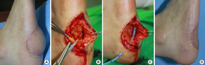
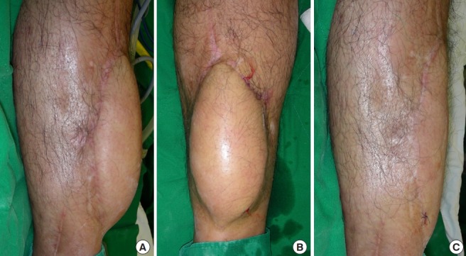
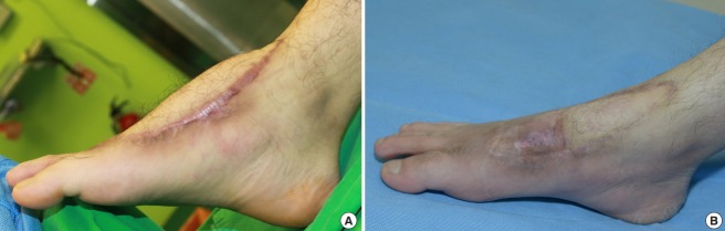
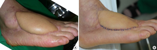
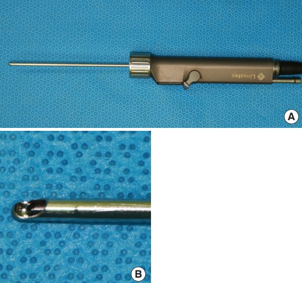
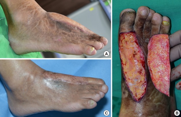
Perforator flaps are becoming increasingly common, and as primary thinning techniques are being developed, the need for secondary contouring of flaps is decreasing. However, many reconstructive flap procedures still incorporate secondary debulking to improve the functional and aesthetic outcomes. Direct excision, liposuction, tissue shaving with an arthroscopic cartilage shaver, and skin grafting are the four major methods used for secondary debulking. Direct excision is primarily applied in flaps where the skin is redundant, even though the volume is not excessive. However, due to the limited range of excision, performing a staged excision is recommended. Liposuction can reduce the amount of subcutaneous tissue of the flap and protect the vascular pedicles. However, the main drawback of this method is its limited ability to remove fibrotic tissues, for which the use of a shaver may be more convenient. The main drawback of using a shaver is that it is difficult to simultaneously remove excess skin. Skin grafting enables the removal of sufficient excess tissue to recover the contour of the normal limb and to improve the color match, facilitating excellent aesthetic results.

Keywords: Contouring; Lipectomy; Perforator flap; Reoperation; Surgical flaps.


Conflict of interest statement

No potential conflict of interest relevant to this article was reported.

Figures

Fig. 1.
Fig. 1.. Contouring of flap by direct excision
A patient underwent an anterolateral thigh (ALT) free flap operation to cover an open wound caused by the excision of malignant melanoma on the right heel. (A) Gross picture 6 months after performing the ALT free flap operation. The patient complained of discomfort wearing shoes due to the bulky flap, as well as aesthetic dissatisfaction. (B) Direct excision was performed for secondary debulking. The flap was excised along the long axis and subcutaneous tissue was directly excised. (C) Sufficient subcutaneous tissue was removed to improve the contour of the flap, leaving 5 mm of the tissue just under the flap to prevent necrosis and depression of the skin. (D) Gross picture 1.5 months after the secondary debulking. The patient no longer complained of functional difficulties and the area looked aesthetically satisfactory.
Fig. 2.
Fig. 2.. Injection of tumescent solution prior to liposuction
A patient underwent an anterolateral thigh (ALT) free flap operation to cover an open fracture on the right tibia. (A) Gross picture 6 months after the ALT free flap operation was performed. The bulky flap did not meet aesthetic standards. (B) Picture taken after the injection of tumescent fluid around the flap for secondary debulking using liposuction. The fluid must be injected evenly throughout the subcutaneous fat layer of the flap, until the contour of the flap starts hardening and the color of the skin starts blanching. (C) Gross picture after secondary debulking was performed, showing an aesthetically excellent result.
Fig. 3.
Fig. 3.. Excessive liposuction may cause skin necrosis
A patient underwent a superficial circumflex iliac artery perforator (SCIP) free flap operation to cover an open wound on the right foot dorsum caused by a burn. (A) Gross picture taken 6 months after the SCIP free flap operation. The bulky flap limited dorsiflexion and caused discomfort wearing shoes. (B) Gross picture taken 6 months after secondary debulking using power-assisted liposuction. Partial skin necrosis was seen after the debulking procedure, but recovered without an additional procedure.
Fig. 4.
Fig. 4.. Flap contouring by ultrasound-assisted liposuction
A patient underwent anterolateral thigh (ALT) free flap surgery to cover an open wound that occurred due to skin necrosis on the right foot dorsum after cellulitis. (A) Gross picture taken 6 months after ALT free flap surgery. The patient complained of discomfort when wearing shoes due to the bulky flap. (B) Gross picture 15 days after secondary debulking using ultrasound-assisted liposuction. It was safe to excise only one side of the long axis after liposuction.
Fig. 5.
Fig. 5.. Arthroscopic cartilage shaver
Picture of an arthroscopic cartilage shaver used in secondary debulking procedures. (A) The hand piece of the arthroscopic cartilage shaver. (B) Magnified picture of the tip.
Fig. 6.
Fig. 6.. Flap contouring by full-thickness skin graft
A patient underwent an anterolateral thigh (ALT) free flap procedure to cover an open wound caused by skin necrosis on the right foot dorsum after trauma. (A) Gross picture taken 6 months after ALT free flap surgery. The patient complained of discomfort when wearing shoes due to the bulky flap. (B) Debulking was performed after harvesting full-thickness skin from the flap. The vascularity of the recipient bed was confirmed by checking for the presence of numerous pinpoint bleeding spots on the debulked flap area. (C) Gross picture taken 6 months after secondary debulking using a fullthickness skin graft. The contouring can be seen to have improved, and the patient no longer complained of functional discomfort.

References

    1. Kim TG, Hong JP, Chung YK. Clinical experience of countouring fasciocutaneous flap using ultrasound assisted liposuction. J Korean Microsurg Soc. 2003;12:99–104.
    1. Hallock GG. Defatting of flaps by means of suction-assisted lipectomy. Plast Reconstr Surg. 1985;76:948–52. - PubMed
    1. Karakullukcu B, van Laarhoven CM, Smeele LE, et al. Functional and aesthetic recontouring of free flap reconstructions of the head and neck region with microdebrider. Kulak Burun Bogaz Ihtis Derg. 2014;24:118–22. - PubMed
    1. Wooden WA, Shestak KC, Newton ED, et al. Liposuction-assisted revision and recontouring of free microvascular tissue transfers. Aesthetic Plast Surg. 1993;17:103–7. - PubMed
    1. Ibrahim AE, Janom H, Raad M. Liposuction contouring after head and neck free flap reconstruction. Anaplastology. 2015;4:145.