. 2018 Aug 30;8(9):3069-3081.
doi: 10.1534/g3.118.200449.

Physiological Starvation Promotes Caenorhabditis elegans Vulval Induction

Affiliations

Physiological Starvation Promotes Caenorhabditis elegans Vulval Induction

Stéphanie Grimbert et al. G3 (Bethesda). .

Abstract

Studying how molecular pathways respond to ecologically relevant environmental variation is fundamental to understand organismal development and its evolution. Here we characterize how starvation modulates Caenorhabditis elegans vulval cell fate patterning - an environmentally sensitive process, with a nevertheless robust output. Past research has shown many vulval mutants affecting EGF-Ras-MAPK, Delta-Notch and Wnt pathways to be suppressed by environmental factors, such as starvation. Here we aimed to resolve previous, seemingly contradictory, observations on how starvation modulates levels of vulval induction. Using the strong starvation suppression of the Vulvaless phenotype of lin-3/egf reduction-of-function mutations as an experimental paradigm, we first tested for a possible involvement of the sensory system in relaying starvation signals to affect vulval induction: mutation of various sensory inputs, DAF-2/Insulin or DAF-7/TGF-β signaling did not abolish lin-3(rf) starvation suppression. In contrast, nutrient deprivation induced by mutation of the intestinal peptide transporter gene pept-1 or the TOR pathway component rsks-1 (the ortholog of mammalian P70S6K) very strongly suppressed lin-3(rf) mutant phenotypes. Therefore, physiologically starved animals induced by these mutations tightly recapitulated the effects of external starvation on vulval induction. While both starvation and pept-1 RNAi were sufficient to increase Ras and Notch pathway activities in vulval cells, the highly penetrant Vulvaless phenotype of a tissue-specific null allele of lin-3 was not suppressed by either condition. This and additional results indicate that partial lin-3 expression is required for starvation to affect vulval induction. These results suggest a cross-talk between nutrient deprivation, TOR-S6K and EGF-Ras-MAPK signaling during C. elegans vulval induction.

Keywords: Caenorhabditis; LIN-3; TOR-S6K; mutational penetrance; vulval cell fate patterning.


Figures

Figure 1
Figure 1
Progression and environmental sensitivity of C. elegans vulval cell fate patterning. (A) A subset of six Pn.p cells (P3.p to P8.p) acquires competence to form vulval tissue through the expression of LIN-39; however, usually only P5.p, P6.p and P7.p adopt vulval cell fates in a stereotypical 2°-1°-2° pattern. The remaining competent cells, P4.p and P8.p, adopt a non-vulval 3° fate, while P3.p may either adopt a 3° fate or a 4° fate, also referred to as F(fused) fate. The induction of vulval cell fates is generated by the cross-talk of EGF-Ras-MAPK and Delta-Notch signaling pathways. Distinct vulval cell divisions according to cell fate occur during the L3 stage and generate 22 vulval cells. (B) The anchor cell (AC) releases the LIN-3 inductive signal. LIN-3 acts as a morphogen, with P6.p receiving the highest level of LIN-3 causing it to adopt a 1° Cell fate (blue). The expression of the Delta ligands in P6.p activates the Delta-Notch pathway in its neighbors, P5.p and P7.p. This activation causes them to adopt a 2° fate (red) and represses the primary fate. A switch from the canonical LET-60/Ras-LIN-45/Raf pathway to a LET-60/Ras-RGL-1-RAL-1 signaling pathway can also promote the 2° Cell fate in P5.p and P7.p (not detailed). (C) Schematic representation of previously reported environmental and physiological inputs affecting C. elegans vulval induction (for details, see introduction). For effects of high temperature on vulval induction, note that Braendle and Félix (2008) found that 25°C culture had a positive effect on vulval induction (e.g., suppression of diverse Vul mutants) whereas Grimbert and Braendle (2014) found that 30°C consistently caused hypoinduced wild type animals.
Figure 2
Figure 2
Quantifying starvation effects on vulval induction in lin-3/egf(rf) mutants. (A) Differences in vulval induction of lin-3(n378) animals exposed to starvation (48 hr) at different developmental stages. Starvation suppression of lin-3(n378) differed significantly between exposed developmental stages (ANOVA, F5,239 = 14.02, P < 0.0001), with strongest effects observed from mid L2 to early L3 stages. (B) Effects of starvation duration on vulval induction of lin-3(n378). Animals (age-synchronized by egg-laying windows) were exposed to starvation at the mid L2 stage for different time periods except for the first treatment where animals were starved in liquid (M9 buffer) for 12 hr directly after hatching (N = 80 per treatment). Starvation duration significantly affected levels of vulval induction (ANOVA, F5,239 = 14.02, P < 0.0001) with strongest suppression of lin-3(n378) at 48 to 120 hr of starvation. (C) Starvation (48 hr) significantly increased vulval induction of lin-3(n378) animals relative to control food conditions (ANOVA, F1,66 = 33.75, P < 0.0001). (D, E) Individual lin-3(n378) fate patterns of P4.p to P8.p in (D) food vs. (E) starvation conditions. (F) Starvation (48 hr) significantly increased vulval induction of lin-3(e1417) animals relative to control food conditions (ANOVA, F1,66 = 9.97, P = 0.0024). (G, H) Individual lin-3(e1417) fate patterns of P4.p to P8.p in (G) food vs. (H) starvation conditions. (I) clk-1(e2519); lin-3(e1417) animals did not show increased vulval induction (in food conditions) despite a strongly prolonged developmental time; rather, the double mutant exhibited a significantly reduced mean number of vulval cells compared to the single mutant lin-3(e1417) in standard food conditions (One-Way-ANOVA, F1,71 = 0.83, P = 0.0025). Values represented by bars (A,B,C,F) indicate the mean number of induced vulval cells, also referred to as the vulval index (WT = 3 cells induced). Values with different letters indicate significant differences (ANOVA, followed by Tukey’s HSD) (* P < 0.05, ** P < 0.01, *** P < 0.001, ns: non-significant). Numbers displayed in bars represent the number of individuals scored; error bars indicate ± 1 SEM. Vulval cell fate patterns of P4.p to P8.p (D,E,G,H) were, whenever feasible, separately inferred for Pn.pa and Pn.pp in cases of half-induced fates. Each line represents the vulval pattern of a single individual, and individuals are ordered from highest to lowest index of vulval induction. Color coding of vulval cell fates (1°: blue, 2°: red) and non-vulval cell fates (3°: yellow). Induced vulval cells that could not be clearly assigned to either a 1° or 2° fate are shown in purple.
Figure 3
Figure 3
Nutrient-deprivation caused by rsks-1 and pept-1 mutations strongly increases vulval induction in lin-3/egf(rf). (A) Effects of let-363, daf-15, rsks-1 and pept-1 RNAi vs. control RNAi treatment (empty vector strain, E. coli HT115) of lin-3(n378) animals (food conditions). RNAi knock-down of let-363, rsks-1 and pept-1 (but not daf-15) significantly increased vulval induction (ANOVA, all P < 0.01). (B) rsks-1(ok1255) strongly increased vulval induction of lin-3(n378) animals (ANOVA, F1,115 = 48.42, P < 0.0001). (C) pept-1(lg601) strongly increased vulval induction of lin-3(n378) animals (ANOVA, F1,118 = 335.58, P < 0.0001). (D, E) Individual fate patterns of P4.p to P8.p in (D) lin-3(n378) vs. (E) lin-3(n378); pept-1(lg601) individuals. Vulval cell fate patterns of P4.p to P8.p (D,E) were, whenever feasible, separately inferred for Pn.pa and Pn.pp in cases of half-induced fates. Each line represents the vulval pattern of a single individual, and individuals are ordered from highest to lowest index of vulval induction. Color coding of vulval cell fates (1°: blue, 2°: red) and non-vulval cell fates (3°: yellow). Induced vulval cells that could not be clearly assigned to either a 1° or 2° fate are coded in purple. (F) Effects of pept-1 RNAi vs. control RNAi (empty vector strain, E. coli HT115) in daf-2(e1370); lin-3(n378) (food conditions). RNAi knock-down of pept-1 significantly increased vulval induction (ANOVA, F1,73 = 133.57, P < 0.0001). (G) 1) Starvation significantly increased vulval induction of rsks-1(ok1255); lin-3(n378) animals relative to control food conditions (ANOVA, F1,102= 11.62, P = 0.0009) 2) Effects of pept-1 RNAi vs. control RNAi (empty vector strain, E. coli HT115) in rsks-1(ok1255); lin-3(n378) (food conditions). RNAi knock-down of pept-1 significantly increased vulval induction (ANOVA, F1,79 = 25.93, P < 0.0001).
Figure 4
Figure 4
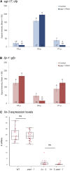
Reduced activity of PEPT-1 increases Ras and Notch pathway activities but does not alter lin-3 transcript levels. (A,B) Effects of pept-1 RNAi on the level of the transcriptional reporters egl-17::cfp (EGF-Ras-MAPK activity) (Yoo et al. 2004) and lip-1::gfp (Delta-Notch activity) (Berset et al. 2001) quantified in lethargus L2/L3 and early L3 stages. (A) Mean signal (pixel) intensity of the EGF-Ras-MAPK pathway reporter, egl-17::cfp in P5.p to P7.p. Values indicate Least Square Means for the interaction cell x RNAi treatment (F2,62 = 15.52, P < 0.0001) (Table 2A). (B) Mean signal (pixel) intensity of the Delta-Notch pathway reporter, lip-1::gfp in P5.p to P7.p. Values indicate Least Square Means for the interaction cell x RNAi treatment (F2,52 = 13.73, P < 0.0001) (Table 2B). For complete statistical analysis and results, see Table 2. Values with different letters indicate significant differences (Tukey’s HSD). Numbers displayed in bars represent the number of individuals scored; error bars indicate ± 1 SEM (C) Boxplot showing lin-3 mRNA levels in different genetic backgrounds [wild type, pept-1(lg601), lin-3(e1417) and lin-3(e1417); pept-1(lg601)] is displayed. The pept-1 mutation does not alter the lin-3 expression level (mean = 24.38 ± 0.67; n = 34) compared to the wild type (mean = 25.36 ± 0.35; n = 22) (ANOVA, F1,54 = 0.49, P = 0.49). As expected, there is little expression in lin-3(e1417) mutant animals (Mean: 0.77 ± 0.35, n = 31) but this low expression is not rescued in lin-3; pept-1 double mutant animals (Mean: 0.36 ± 0.17; n = 22) (ANOVA, F1,751= 0.46, P = 0.50).
Figure 5
Figure 5
Role of BAR-1 activity in starvation modulation of vulval induction. (A) Differences in vulval induction of bar-1(ga80) animals exposed to 48 hr of starvation in late L1 vs. mid L2 (and vs. control food conditions). Starvation at the late L1 stage, but not at the mid L2 stage, significantly reduced vulval induction of bar-1(ga80) animals (ANOVA, F2,121 = 50.27, P < 0.0001). (B-D) Individual bar-1(ga80) fate patterns of P3.p to P8.p in (B) food vs. starvation conditions: late L1 (C) or mid L2 starvation (D). Vulval cell fate patterns of P3.p to P8.p (B-D) were, whenever feasible, separately inferred for Pn.pa and Pn.pp in cases of half-induced fates. Each line represents the vulval pattern of a single individual, and individuals are ordered from highest to lowest index of vulval induction. Color coding of vulval cell fates (1°: blue, 2°: red) and non-vulval cell fates (3°: yellow, 4°: gray). Induced vulval cells that could not be clearly assigned to either a 1° or 2° fate are coded in purple; non-induced cells that could not be clearly assigned a 3° or° fate are coded in white. (E) Proportion of Pn.p cells with a 4° (Fused) fate in control, starvation-exposed late L1, starvation-exposed mid L2 bar-1(ga80) individuals (from the same experiment as shown in (A)). The proportions of 4° fates for each of P5.p to P7.p cell were higher after L1 starvation (P5.p: 37%, P6.p: 23%, P7.p: 44%) compared to control (P5.p: 2%, P6.p and P7.p: 0%) and L2 starvation (P5.p: 12%, P6.p: 0%, P7.p: 3%). (F) pept-1 RNAi increases vulval induction of bar-1(ga80) animals (ANOVA, F1,81 = 14.47, P = 0.0003). (G) Starvation increased vulval induction of lin-3(n378); bar-1(mu63) relative to control food conditions (ANOVA, F1,98 = 18.84, P < 0.0001). (H) Starvation increased vulval induction of egl-30(ad805); lin-3(n378) relative to control food conditions (ANOVA, F1,59 = 7.28, P = 0.0091).
Figure 6
Figure 6
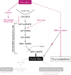
Starvation modulation of C. elegans vulval induction. Schematic representation of (antagonistic) starvation effects reported in this and earlier studies (Battu et al. 2003; Braendle and Félix 2008; Ferguson and Horvitz 1985).

References

    1. Antebi A., Yeh W. H., Tait D., Hedgecock E. M., Riddle D. L., 2000. daf-12 encodes a nuclear receptor that regulates the dauer diapause and developmental age in C. elegans. Genes Dev. 14: 1512–1527. - PMC - PubMed
    1. Barkoulas M., Van Zon J. S., Milloz J., Van Oudenaarden A., Félix M.-A., 2013. Robustness and epistasis in the C. elegans vulval signaling network revealed by pathway dosage modulation. Dev. Cell 24: 64–75. 10.1016/j.devcel.2012.12.001 - DOI - PubMed
    1. Barkoulas M., Vargas Velazquez A. M., Peluffo A. E., Felix M. A., 2016. Evolution of New cis-Regulatory Motifs Required for Cell-Specific Gene Expression in Caenorhabditis. PLoS Genet. 12: e1006278 10.1371/journal.pgen.1006278 - DOI - PMC - PubMed
    1. Battu G., Hoier E. F., Hajnal A., 2003. The C. elegans G-protein-coupled receptor SRA-13 inhibits RAS/MAPK signalling during olfaction and vulval development. Development 130: 2567–2577. 10.1242/dev.00497 - DOI - PubMed
    1. Berset T., Hoier E. F., Battu G., Canevascini S., Hajnal A., 2001. Notch inhibition of RAS signaling through MAP kinase phosphatase LIP-1 during C. elegans vulval development. Science 291: 1055–1058. 10.1126/science.1055642 - DOI - PubMed

Publication types

Substances

LinkOut - more resources