. 2018 Dec;16(12):1889-1901.
doi: 10.1158/1541-7786.MCR-18-0345. Epub 2018 Jul 23.

Molecular Portrait of Hypoxia in Breast Cancer: A Prognostic Signature and Novel HIF-Regulated Genes

Affiliations

Molecular Portrait of Hypoxia in Breast Cancer: A Prognostic Signature and Novel HIF-Regulated Genes

I Chae Ye et al. Mol Cancer Res. 2018 Dec.

Abstract

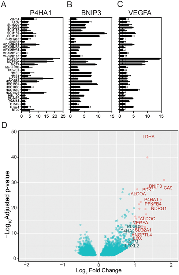
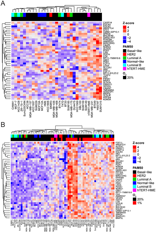
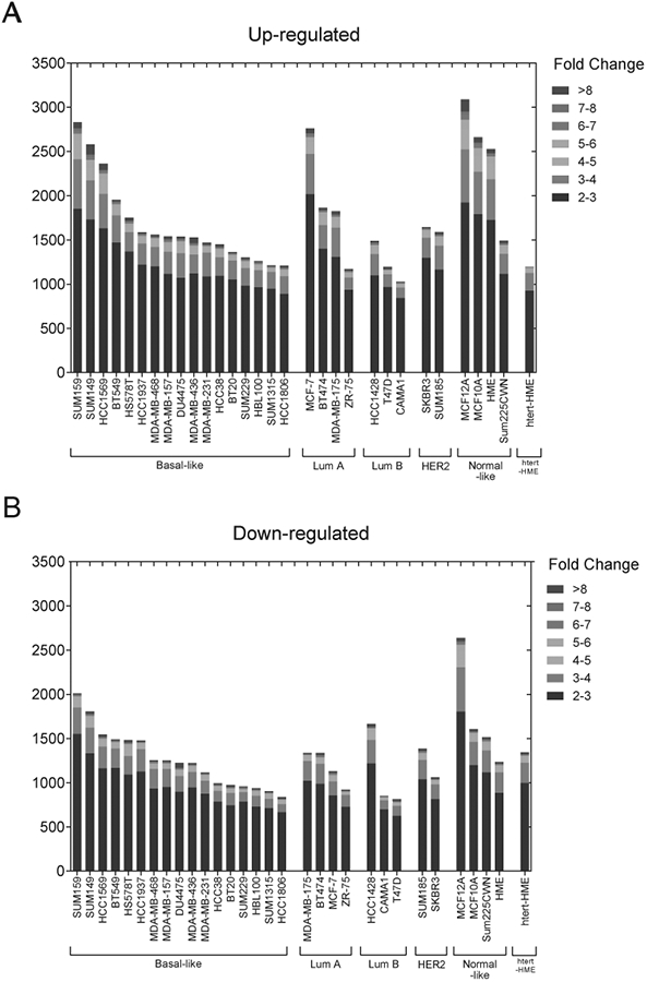
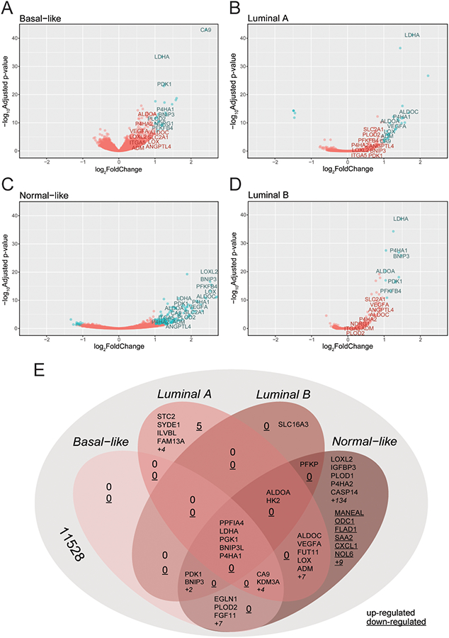
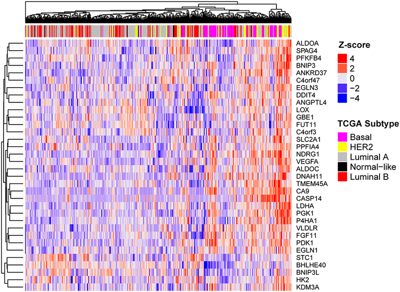
Intratumoral hypoxia has been associated with invasion, metastasis, and treatment failure, prompting the need for a global characterization of the response to hypoxic conditions. The current study presents the results of a large-scale RNA sequencing (RNA-seq) effort, analyzing 31 breast cancer cell lines representative of breast cancer subtypes or normal mammary epithelial (NME) cells exposed to control tissue culture conditions (20% O2) or hypoxic conditions (1% O2). The results demonstrate that NME have a stronger response to hypoxia both in terms of number of genes induced by hypoxia as well as level of expression. A conserved 42-gene hypoxia signature shared across PAM50 subtypes and genes that are exclusively upregulated in Luminal A, Luminal B, and normal-like mammary epithelial cells is identified. The 42-gene expression signature is enriched in a subset of basal-like cell lines and tumors and differentiates survival among patients with basal-like tumors. Mechanistically, the hypoxia-inducible factors (HIF-1 and/or HIF-2) mediate the conserved hypoxic response. Also, four novel hypoxia-regulated and HIF-1-responsive genes were identified as part of the conserved signature. This dataset provides a novel resource to query transcriptional changes that occur in response to hypoxia and serves as a starting point for a clinical assay to aid in stratifying patients that would benefit from hypoxia-targeted therapies, some of which are currently in clinical trials. IMPLICATIONS: RNA-seq of 31 breast cancer cells exposed to control or hypoxic conditions reveals a conserved genomic signature that contains novel HIF-regulated genes and is prognostic for the survival of patients with triple-negative breast cancer.


Conflict of interest statement

The authors declare no conflict of interest.

Figures

Figure 1.
Figure 1.
RNA sequencing of 31 cells line exposed to 20% O2 and 1% O2 reveals a conserved set of hypoxia-inducible genes. A-C, qPCR of RNA lysates from 34 breast cancer cells lines to determine the fold change in the induction of P4HA1 (A) BNIP3 (B) and VEGFA (C) upon exposure to 1% compared to 20% O2 conditions (N = 3 biological × 3 technical repeats per cell line). Dash lines mark a fold change equal to 1 (left) or 2 (right). D, Differential expression statistics comparing samples exposed to 20% as compared with 1% O2 were calculated using DESeq2. A volcano plot of the differential expression statistics show genes (in red) with a log-fold change (LFC) greater than 1 and a false-discovery rate (FDR) adjusted p-value less than 0.05. The 42-genes that meet this criteria are listed in Supplementary Table S2.
Figure 2.
Figure 2.
Basal-like breast cancer cells express higher endogenous levels of the 42-gene hypoxia signature. A-B, Heatmap of gene expression values for the 42-gene hypoxia signature identified with CoGAPS analysis of gene expression data from 31 breast cancer cell lines cultured under standard tissue culture conditions (20% O2) (A) or both 20% and 1% O2 (B) Color-coding describes the subtype of the cancer cell line. htert-HME cells were not classified in the Marcotte et al. study. The z-score ranges from low (blue) to high (red).
Figure 3.
Figure 3.
Breast cancer cells display a heterogeneous response to hypoxic conditions. A-B, The number of genes that are upregulated (A) or downregulated (B) in each cell line stratified by fold change after exposure to 1% compared to 20% O2 conditions.
Figure 4.
Figure 4.
Basal cell lines display the highest number of differentially expressed genes upon exposure to hypoxia. A-D, Differential expression statistics comparing samples exposed to 20% as compared with 1% O2 were calculated using DESeq2 for Basal-like (A), Luminal A (B), Normal-like (C) and Luminal B (D) subtype cell lines. A volcano plot of the differential expression statistics show genes (in red) with a LFC greater than 1 and FDR adjusted p-value less than 0.05. E, VENN diagram displaying the number of genes that overlap or that are exclusive to each subtype.
Figure 5.
Figure 5.
Patients with Basal breast cancer have a higher level of the 42-gene hypoxia signature. Supervised clustering analysis of all breast cancer patients in the TCGA database by the 42-gene hypoxia signature reveals that patients with basal breast cancer have a higher level of expression of hypoxia gene compared to other subtypes.
Figure 6.
Figure 6.
Novel hypoxia regulated identified in the HIF-signature require HIF-1α expression. A, qPCR to determine CASP14, FUT11, DNAH11, and TCAF2 mRNA levels in 12 breast cancer cell lines exposed to 20% O2 or 1% O2 for 24 h, normalized to mean value for HCC1569 cells at 20% O2 (for CASP14, FUT11, and TCAF2) or HME cells at 20% O2 (for DNAH11) (mean ± SEM, n = 3). *, P < 0.05 versus 20% O2 within the same cell line (Student’s t-test). B-C, CASP14 and FUT11 (B) or DNAH11 and TCAF2 (C) mRNA levels were analyzed by qPCR in MCF7 (B) or MDA-MB-231 (C) subclones, which were stably transfected with an non-target control (NTC) CRISPR vector or vectors encoding gDNA sequences that target either HIF-1α (1–1, 1–2) or HIF-2α (2–1, 2–2), and exposed to 20% or 1% O2 for 24 h (mean ± SEM, n = 3–6); **, P < 0.01, ***, P < 0.001 versus NTC at 20% O2 or ###, P < .001 versus NTC at 1% O2 (two-way ANOVA with Bonferroni posttest). D, Candidate HIF-binding sites (ovals) were identified in the 5’-untranslated region of the human CASP14 gene (exons 1 and 2 shown as a rectangles). E, BT474 cells were incubated at 20% or 1% O2 for 4 hours and ChIP assays were performed using IgG or antibodies against HIF-1α, HIF-2α, or HIF-1β. Specific primers flanking candidate HIF-binding sites were used for qPCR (Supplementary Table S1), and values were normalized to IgG expression at 20% O2 (mean ± SEM, n = 3). *, P < 0.05 vs. 20% O2 within the same antibody (Student’s t-test).

References

    1. Dewhirst MW. Intermittent hypoxia furthers the rationale for hypoxia-inducible factor-1 targeting. Cancer Res 2007;67(3):854–5. - PubMed
    1. Brahimi-Horn MC, Chiche J, Pouyssegur J. Hypoxia and cancer. J Mol Med 2007;85(12):1301–7. - PubMed
    1. Semenza GL, Ruvolo PP. Introduction to tumor microenvironment regulation of cancer cell survival, metastasis, inflammation, and immune surveillance. Biochim Biophys Acta 2016;1863(3):379–81. - PubMed
    1. Gillies RJ, Gatenby RA. Hypoxia and adaptive landscapes in the evolution of carcinogenesis. Cancer Metastasis Rev 2007;26(2):311–7. - PubMed
    1. Bos R, van der Groep P, Greijer AE, Shvarts A, Meijer S, Pinedo HM, et al. Levels of hypoxia-inducible factor-1α independently predict prognosis in patients with lymph node negative breast carcinoma. Cancer 2003;97(6):1573–81. - PubMed

Publication types

Substances