. 2018 Aug 7;115(32):8197-8202.
doi: 10.1073/pnas.1802326115. Epub 2018 Jul 23.

RNA triphosphatase DUSP11 enables exonuclease XRN-mediated restriction of hepatitis C virus

Affiliations

RNA triphosphatase DUSP11 enables exonuclease XRN-mediated restriction of hepatitis C virus

Rodney P Kincaid et al. Proc Natl Acad Sci U S A. .

Abstract

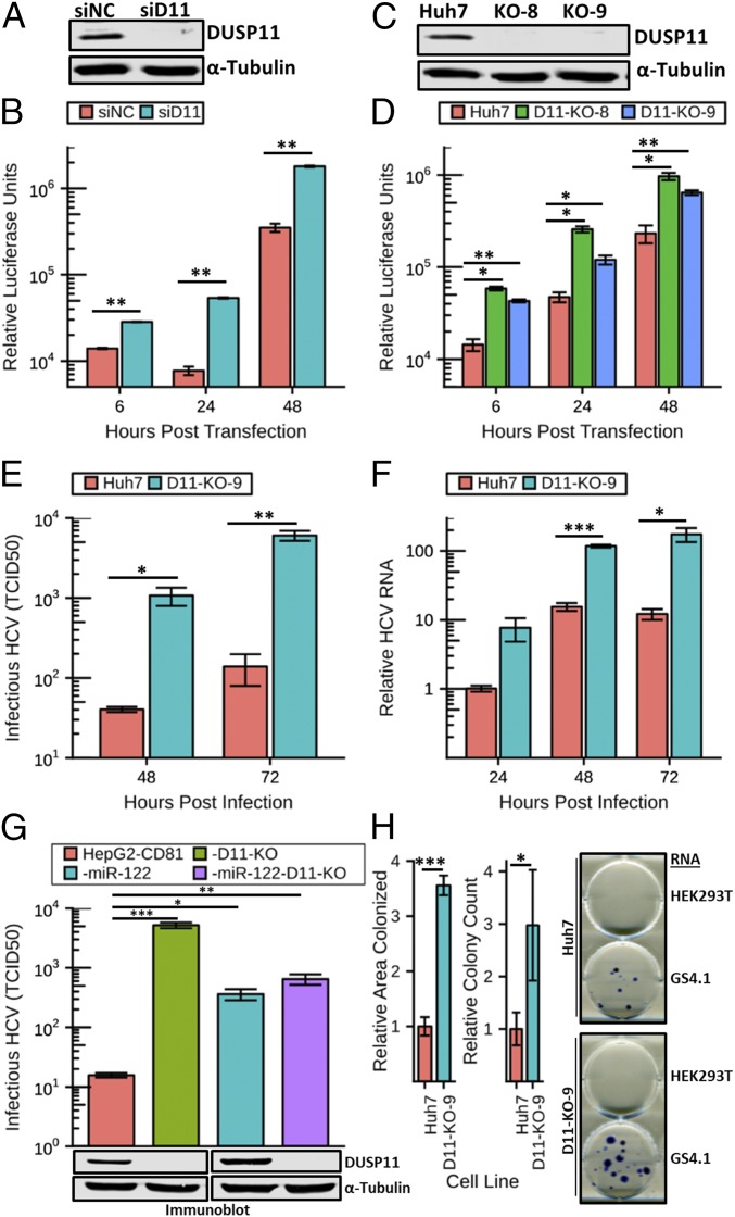
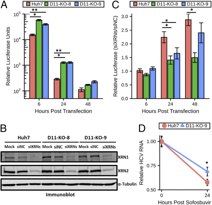
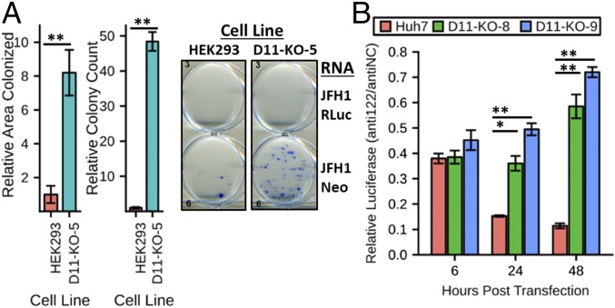
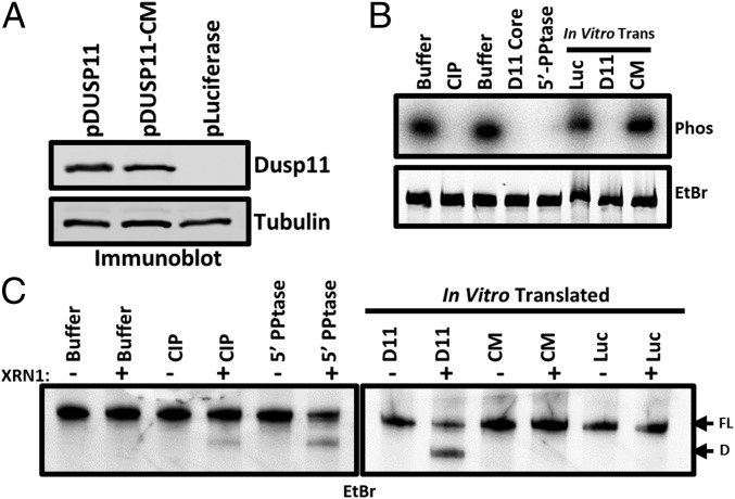
Seventy percent of people infected with hepatitis C virus (HCV) will suffer chronic infection, putting them at risk for liver disease, including hepatocellular carcinoma. The full range of mechanisms that render some people more susceptible to chronic infection and liver disease is still being elucidated. XRN exonucleases can restrict HCV replication and may help to resolve HCV infections. However, it is unknown how 5' triphosphorylated HCV transcripts, primary products of the viral polymerase, become susceptible to attack by 5' monophosphate-specific XRNs. Here, we show that the 5' RNA triphosphatase DUSP11 acts on HCV transcripts, rendering them susceptible to XRN-mediated attack. Cells lacking DUSP11 show substantially enhanced HCV replication, and this effect is diminished when XRN expression is reduced. MicroRNA-122 (miR-122), a target of current phase II anti-HCV drugs, is known to protect HCV transcripts against XRNs. We show that HCV replication is less dependent on miR-122 in cells lacking DUSP11. Combined, these results implicate DUSP11 as an important component of XRN-mediated restriction of HCV.

Keywords: miR-122; microRNA; restriction factor.


Conflict of interest statement

The authors declare no conflict of interest.

Figures

Fig. 1.
Fig. 1.
Reducing DUSP11 levels enhances HCV replication and infection. (A) Confirmation of siRNA knockdown of DUSP11 in Huh7 cells by immunoblot. Huh7 were transfected with either an irrelevant control siRNA (siNC) or DUSP11-specific siRNA (siD11). Cell lysates were harvested 48 h post transfection and assayed by immunoblot with the indicated antibodies. (B) sgJFH1-Rluc replicon luciferase assay in Huh7 cells treated with siNC or siD11. Luciferase assays were performed at the indicated times post replicon RNA transfection. The mean ± SEM of three individual experiments is presented. (C) Confirmation of CRISPR/Cas9 disruption of DUSP11 in Huh7 cells by immunoblot. Lysates from parental Huh7 cells and two independent DUSP11 knockout clones (D11-KO-8 and D11-KO-9) were assayed by immunoblot with the indicated antibodies. (D) sgJFH1-Rluc replicon luciferase assay in parental Huh7 cells and two independent DUSP11 knockout clones (D11-KO-8 and D11-KO-9). Luciferase assays were performed at the indicated times post replicon RNA transfection. The mean ± SEM of three individual experiments is presented. (E) HCV cell culture (HCVcc) infectious virus production [multiplicity of infection (MOI) = 1] in Huh7 and D11-KO-9 cells. Data presented are the mean of three replicates ± SEM. (F) HCVcc RNA replication time course (MOI = 1) in Huh7 and D11-KO-9 cells. Data presented are the mean of three replicates ± SEM. (G) HCVcc infectious virus production (MOI = 10) in HepG2-CD81, HepG2-CD81-D11, CD81-miR-122, and HepG2-CD81-miR-122-D11. Data presented are the mean of three replicates ± SEM. (G, Lower) Immunoblot of lysates from the corresponding cell lines with the indicated antibodies. (H) GS4.1 replicon colony formation assays in parental Huh7 cells and DUSP11 knockout cells (D11-KO-9). Quantitation of both relative area colonized and relative colony counts with transfection of GS4.1 RNA is presented (Left). The mean ± SEM of five individual experiments is presented. Example wells with stained colonies from the indicated cell lines transfected with the indicated RNA are displayed (Right). Statistical significance was assessed by Student’s t test and is indicated as follows: *P < 0.05, **P < 0.01, ***P < 0.001.
Fig. 2.
Fig. 2.
HCV restriction activity of XRNs is reduced in cells lacking DUSP11. (A) sgJFH1-Rluc-GND (nonreplicative polymerase mutant) replicon luciferase assay in parental Huh7 cells and two independent DUSP11 knockout clones (D11-KO-8 and D11-KO-9). Luciferase assays were performed at the indicated times post replicon RNA transfection. The mean ± SEM of three individual experiments is presented. (B) Confirmation of siRNA knockdown of XRN1 and XRN2 in parental Huh7 cells and two independent DUSP11 knockout clones (D11-KO-8 and D11-KO-9) by immunoblot. Huh7 were either mock transfected (Mock) or transfected with either an irrelevant control siRNA or a pool of XRN1- and XRN2-specific siRNAs (siXRNs). Cell lysates were harvested 48 h post transfection and assayed by immunoblot with the indicated antibodies. (C) sgJFH1-Rluc replicon luciferase assay in parental Huh7 cells and two independent DUSP11 knockout clones (D11-KO-8 and D11-KO-9) treated with siNC or a pool of siXRNs. Luciferase assays were performed at the indicated times post replicon RNA transfection. The mean ± SEM of three experiments is presented. (D) Huh7 and D11-KO-9 cells were infected with HCV for 96 h and treated with 5 μM sofosbuvir, and HCV RNA levels were quantified at the indicated times post treatment. The mean ± SEM of eight samples is presented. Statistical significance was assessed by Student’s t test and is indicated as follows: *P < 0.05, **P < 0.01.
Fig. 3.
Fig. 3.
HCV genome replication is less dependent on miR-122 in cells lacking DUSP11. (A) sgJFH1-Neo replicon colony formation assays in parental HEK293 cells and DUSP11 knockout cells (D11-KO-5). Quantitation of both the relative area colonized and relative colony counts with transfection of sgJFH1-Neo RNA is presented (Left). The mean ± SEM of three individual experiments is presented. Example wells with stained colonies from the indicated cell lines transfected with the indicated RNA are displayed (Right). (B) sgJFH1-Rluc replicon luciferase assay in parental Huh7 cells and two independent DUSP11 knockout clones (D11-KO-8 and D11-KO-9) treated with irrelevant control anti-miR (antiNC) or miR-122–specific anti-miR (anti122). Luciferase assays were performed at the indicated times post replicon RNA transfection. The mean of three individual experiments ± SEM is presented. Statistical significance was assessed by Student’s t test and is indicated as follows: *P < 0.05, **P < 0.01.
Fig. 4.
Fig. 4.
DUSP11 directly dephosphorylates HCV 5′ UTR RNA and sensitizes it to XRN-mediated degradation. (A) Confirmation of in vitro translation products by immunoblot. Membrane was incubated with the indicated antibodies. “pLuciferase” indicates reactions programmed to express luciferase as a negative control. (B) In vitro phosphatase assay. 5′ γ-32P–radiolabeled HCV 5′ UTR RNA was incubated with the indicated enzymes (calf intestinal phosphatase, purified DUSP11 core protein, and bacterial 5′ RNA polyphosphatase), or in vitro translated products from A (pDUSP11, pDUSP11-CM, and negative-control pLuciferase). Products were separated by urea/PAGE and stained with ethidium bromide. Products were then transferred to a nitrocellulose membrane and exposed to a phosphor storage screen (Phos). (C) In vitro XRN susceptibility assay. In vitro phosphatase reactions were performed as in B [calf intestinal phosphatase, bacterial 5′ RNA polyphosphatase, or in vitro translated products from A (pDUSP11, pDUSP11-CM, and negative-control pLuciferase)], but products were recovered and incubated ± recombinant XRN1. Products were separated by urea/PAGE and stained with EtBr. “FL” arrow points to the position of the full-length HCV 5′ UTR. “D” arrow points to the position of a faster-migrating degradation product.
Fig. 5.
Fig. 5.
Model: DUSP11 converts 5′ triphosphorylated HCV RNAs to 5′ monophosphate substrates for XRNs. This results in reduced HCV RNA levels and reduced virus yield.

References

    1. Kanwal F, et al. Risk of hepatocellular cancer in HCV patients treated with direct-acting antiviral agents. Gastroenterology. 2017;153:996–1005.e1. - PubMed
    1. Zhou L-Y, Zhang L-L. Host restriction factors for hepatitis C virus. World J Gastroenterol. 2016;22:1477–1486. - PMC - PubMed
    1. You H, et al. Novel host genetic variations associated with spontaneous clearance of a single-source outbreak of HCV1b infections. BMJ Open Gastroenterol. 2015;1:e000010. - PMC - PubMed
    1. Thomas DL, et al. Genetic variation in IL28B and spontaneous clearance of hepatitis C virus. Nature. 2009;461:798–801. - PMC - PubMed
    1. Missiha SB, Ostrowski M, Heathcote EJ. Disease progression in chronic hepatitis C: Modifiable and nonmodifiable factors. Gastroenterology. 2008;134:1699–1714. - PubMed

Publication types

MeSH terms

LinkOut - more resources