. 2018 Sep;3(9):1023-1031.
doi: 10.1038/s41564-018-0210-y. Epub 2018 Jul 23.

Murine colitis reveals a disease-associated bacteriophage community

Affiliations

Murine colitis reveals a disease-associated bacteriophage community

Breck A Duerkop et al. Nat Microbiol. 2018 Sep.

Abstract

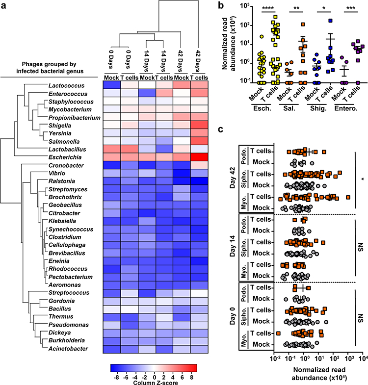
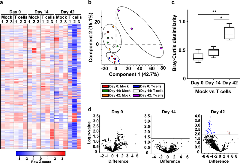
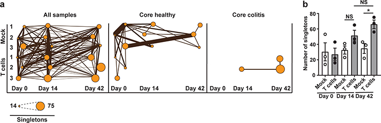
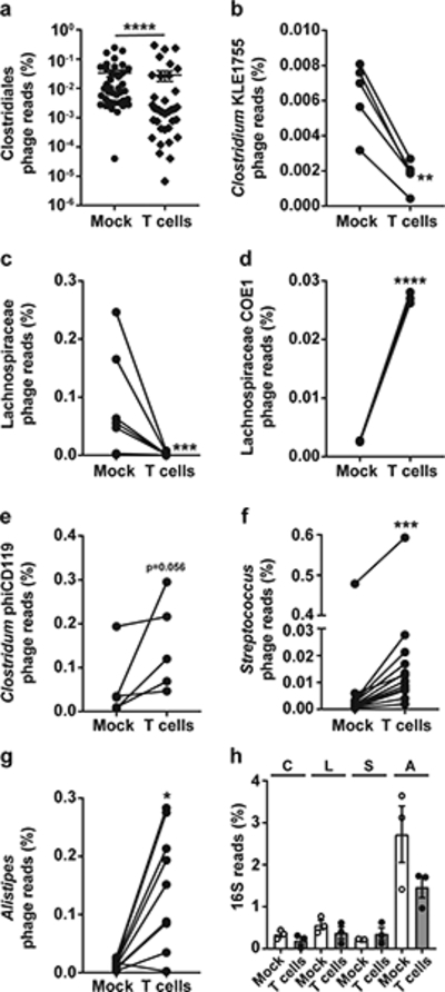
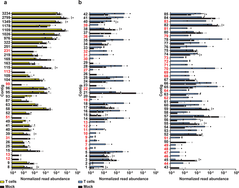
The dysregulation of intestinal microbial communities is associated with inflammatory bowel diseases (IBD). Studies aimed at understanding the contribution of the microbiota to inflammatory diseases have primarily focused on bacteria, yet the intestine harbours a viral component dominated by prokaryotic viruses known as bacteriophages (phages). Phage numbers are elevated at the intestinal mucosal surface and phages increase in abundance during IBD, suggesting that phages play an unidentified role in IBD. We used a sequence-independent approach for the selection of viral contigs and then applied quantitative metagenomics to study intestinal phages in a mouse model of colitis. We discovered that during colitis the intestinal phage population is altered and transitions from an ordered state to a stochastic dysbiosis. We identified phages specific to pathobiotic hosts associated with intestinal disease, whose abundances are altered during colitis. Additionally, phage populations in healthy and diseased mice overlapped with phages from healthy humans and humans with IBD. Our findings indicate that intestinal phage communities are altered during inflammatory disease, establishing a platform for investigating phage involvement in IBD.


Conflict of interest statement

Competing interests

The authors declare no competing interests related to this work.

Figures

Figure 1.
Figure 1.
Caudovirales phage abundances are altered during T cell-mediated colitis. (a) Heat map indicating the abundance of VLP sequencing reads with at least 97% identity to select sequenced Caudovirales phage genomes deposited in NCBI. Relative read abundances were summed by phage-host genus and z-scores were normalized by column. (b) Quantitative read mapping abundances for select groups of phages shown in panel A. Esch. – Escherichia phages, Sal. – Salmonella phages, Shig. – Shigella phages and Entero. – Enterococcus phages. **** p=0.0003, *** p=0.003, ** p=0.02, * p=0.05 by two-tailed nonparametric Mann-Whitney test. (c) Mapped read abundances for the Caudovirales phage families Siphoviridae (Sipho.), Myoviridae (Myo.) and Podoviridae (Podo.) at Days 0, 14 and 42 post T cell transfer. Mock – saline injected control mice, T cells – mice receiving CD4+ CD45RBHi T cells. Z-scores show how many standard deviations a value in a column is above and below the population mean. The data represent three independent mice per group. Error bars represent ± standard error of the mean. * p=0.007 by one-way ANOVA, NS – not significant.
Figure 2.
Figure 2.
Phage community alterations during colitis were identified using a curated VLP contig database. (a) Heat map showing the differential abundance analysis of VLP sequencing reads mapped to a curated database consisting of 1104 VLP contigs during colitis. Numbers 1–3 represent individual mice within each group. Each row representing a unique phage contig was z-score normalized. (b) Principal component analysis showing that phage community abundances are different between healthy and T cell treated animals. Ellipses were manually added to guide the reader and do not indicate confidence intervals. (c) Bray-Curtis dissimilarity β diversity metric quantifying the dissimilarity of the phage communities between healthy and T cell treated animals across the time series. The closer the value is to 1 the more dissimilar the communities are in terms of species composition. The whiskers represent the 5–95 percentile of an all against all comparison with the central line in each box representing the median value of the data set. (d) Volcano plots of two-tailed t-tests testing for changes to viral contig abundances during colitis. The multiple-testing problem was corrected by calculating a permutation-based false discovery rate (FDR). A FDR cutoff of 0.05 was used. Horizontal lines indicate the FDR based p-value cut off for statistical significance for each time point when comparing the contig abundances between healthy and diseased animals. Data points highlighted in blue and red indicate contigs that are significantly reduced or enriched in colitic animals respectively and grey data points indicate contigs for which phage host taxonomic assignments could be made. The data represent three independent mice per group. ** p=0.0001, * p=0.01 by Kruskal-Wallis test and a post hoc Dunn’s test for multiple comparisons.
Figure 3.
Figure 3.
Colitic animals share less VLP connections relative to healthy animals. (a) Connected VLP contigs (both shared and singleton contigs) across all time points and animals, from only healthy animals (Core healthy) which includes animals prior to T cell treatment and T cell treated animals 14 and 42 days post T cell administration (Core colitis). Circle diameter indicates the number of singleton VLP contigs that do not cluster with contigs in IMG/VR or to contigs within the curated database. Line thickness represents the number of shared contigs between animals and time points. (b) Abundance of singleton VLP contigs between mock and T cell treated animals across the time series. The data represent three independent mice per group. Error bars represent ± standard error of the mean. * p=0.02 by two-tailed unpaired Student’s t-test, NS – not significant.
Figure 4.
Figure 4.
Phage taxonomy and host bacterial assignments for curated VLP contigs reveals differential abundances of phages that infect both commensals and pathobionts during colitis. (a) All phage contigs linked to the order Clostridiales. Each data point represents a unique Clostridiales phage contig found in both healthy and diseased animals. (b-e) Family and genus-specific phages constituting lower taxonomic ranks of the Clostridiales. (f-g) Abundances of Streptococcal phages and Alistipes phages. (h) Bacterial 16s rDNA sequencing read abundances for the C – Clostridiales, L – Lachnospiraceae, S – Streptococci and A – Alistipes. In panels b-g, lines connect shared contigs between healthy and diseased animals. The data represent three independent mice per group. Error bars represent ± standard error of the mean. **** p<0.0001, *** p=0.0002, ** p=0.008, *p=0.004 by two-tailed nonparametric Mann-Whitney test.
Figure 5.
Figure 5.
VLP reads from healthy and colitic animals share identity to human associated intestinal phages. (a) VLP reads from healthy (mock) and colitic (T cells) mice 42 days post T cell treatment were mapped against a cohort of conserved phage contigs found in healthy humans. (b) VLP reads from healthy (mock) and colitic (T cells) mice 42 days post T cell treatment were mapped against phage contigs assembled from IBD patients. Panel b shows the top 85 IBD phage contigs with read recruitment. Normalized read abundance is the total number of mapped reads divided by the length of the contig in base pairs. Contigs highlighted in red indicate contigs which are only found in healthy or colitic mice. The data represent three independent mice per group. Error bars represent ± standard error of the mean. * p<0.1 by a two sample unpaired t-test with a permutation-based false discovery rate cutoff of 0.1 (Supplementary Table 10).

Comment in

  • Bacteriophage virome in IBD.
    Thoma C. Thoma C. Nat Rev Gastroenterol Hepatol. 2018 Sep;15(9):520. doi: 10.1038/s41575-018-0056-z. Nat Rev Gastroenterol Hepatol. 2018. PMID: 30076371 No abstract available.

Similar articles

Cited by

References

    1. Abraham C & Cho JH Inflammatory bowel disease. N Engl J Med 361, 2066–2078 (2009). - PMC - PubMed
    1. Molodecky NA et al. Increasing incidence and prevalence of the inflammatory bowel diseases with time, based on systematic review. Gastroenterology 142, 46–54 e42 (2012). - PubMed
    1. Hooper LV, Midtvedt T & Gordon JI How host-microbial interactions shape the nutrient environment of the mammalian intestine. Annu Rev Nutr 22, 283–307 (2002). - PubMed
    1. Barthel M et al. Pretreatment of mice with streptomycin provides a Salmonella enterica serovar Typhimurium colitis model that allows analysis of both pathogen and host. Infect Immun 71, 2839–2858 (2003). - PMC - PubMed
    1. Ivanov II et al. Specific microbiota direct the differentiation of IL-17-producing T-helper cells in the mucosa of the small intestine. Cell Host Microbe 4, 337–349 (2008). - PMC - PubMed

Publication types