Differentiation between metastatic and tumour-free cervical lymph nodes in patients with papillary thyroid carcinoma by grey-scale sonographic texture analysis
- PMID: 30038677
- PMCID: PMC6047085
- DOI: 10.5114/pjr.2018.75017
Differentiation between metastatic and tumour-free cervical lymph nodes in patients with papillary thyroid carcinoma by grey-scale sonographic texture analysis
Abstract
Purpose: Papillary thyroid carcinoma (PTC) is the most common thyroid cancer, and cervical lymph nodes (LNs) are the most common extrathyroid metastatic involvement. Early detection and reliable diagnosis of LNs can lead to improved cure rates and management costs. This study explored the potential of texture analysis for texture-based classification of tumour-free and metastatic cervical LNs of PTC in ultrasound imaging.
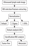
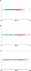
Material and methods: A total of 274 LNs (137 tumour-free and 137 metastatic) were explored using the texture analysis (TA) method. Up to 300 features were extracted for texture analysis in three normalisations (default, 3sigma, and 1-99%). Linear discriminant analysis was employed to transform raw data to lower-dimensional spaces and increase discriminative power. The features were classified by the first nearest neighbour classifier.
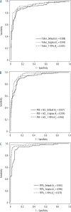
Results: Normalisation reflected improvement on the performance of the classifier; hence, the features under 3sigma normalisation schemes through FFPA (fusion Fisher plus the probability of classification error [POE] + average correlation coefficients [ACC]) features indicated high performance in classifying tumour-free and metastatic LNs with a sensitivity of 99.27%, specificity of 98.54%, accuracy of 98.90%, positive predictive value of 98.55%, and negative predictive value of 99.26%. The area under the receiver operating characteristic curve was 0.996.
Conclusions: TA was determined to be a reliable method with the potential for characterisation. This method can be applied by physicians to differentiate between tumour-free and metastatic LNs in patients with PTC in conventional ultrasound imaging.
Keywords: computer-assisted; diagnosis; lymph nodes; pattern recognition; thyroid carcinoma; ultrasonography.
Figures


References
-
- Howlader N, Noone A, Krapcho M, et al. SEER Cancer Statistics Review, 1975-2013. Secondary SEER Cancer Statistics Review, 1975-2013 based on November 2015 SEER data submission, posted to the SEER web site, April 2016. https://seer.cancer.gov/csr/1975_2013/
-
- Davies L, Welch HG. Current thyroid cancer trends in the United States. JAMA Otolaryngol Head Neck Surg. 2014;140:317–322. - PubMed
-
- Choi YJ, Yun JS, Kook SH, et al. Clinical and imaging assessment of cervical lymph node metastasis in papillary thyroid carcinomas. World J Surg. 2010;34:1494–1499. - PubMed
-
- Haugen BR, Alexander EK, Bible KC, et al. 2015. American Thyroid Association Management Guidelines for Adult Patients with Thyroid Nodules and Differentiated Thyroid Cancer: The American Thyroid Association Guidelines Task Force on Thyroid Nodules and Differentiated Thyroid Cancer. Thyroid. 2016;26:1–133. - PMC - PubMed
LinkOut - more resources
Full Text Sources
Other Literature Sources
