. 2016 Jul 22;100(1):70.
doi: 10.5334/jbr-btr.1118.

Diffusion-Weighted Mri of Postmenopausal Women with Vaginal Bleeding and Endometrial Thickening: Differentiation of Benign and Malignant Lesions

Affiliations

Diffusion-Weighted Mri of Postmenopausal Women with Vaginal Bleeding and Endometrial Thickening: Differentiation of Benign and Malignant Lesions

Mehtap Çavuşoğlu et al. J Belg Soc Radiol. .

Abstract

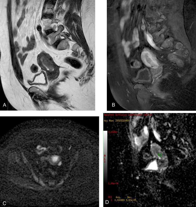
Purpose: To investigate the feasibility of diffusion-weighted magnetic resonance imaging (DWI) with apparent diffusion coefficient (ADC) values in differentiating endometrial cancer from benign endometrial lesions in postmenopausal patients with vaginal bleeding and endometrial thickening and to predict the depth of myometrial invasion in endometrial cancer.

Materials and methods: Postmenopausal patients with vaginal bleeding and endometrial thickening were enrolled in this prospective study. T2-weighted, pre- and postcontrast T1-weighted and diffusion-weighted images were obtained. The ADC values of all the patients with endometrial pathologies were recorded. The staging accuracies of DWI and postcontrast T1-weighted images in the assessment of myometrial invasion were evaluated in histopathologically proven endometrial cancer patients.

Results: Fifty-two patients (mean age: 57 ± 10, range: 41-79) were enrolled in the study. Thirty-eight of the lesions were benign (27 as hyperplasia and endometritis; 11 as polyps). Fourteen of the 52 endometrial lesions were pathologically proven as cancers and underwent hysterectomy, and all the specimens were reported as endometrioid adenocarcinomas. The mean ADC value (10-3 mm2/second) of cancer (0.88 ± 0.10) was significantly lower than that of benign lesions (1.78 ± 0.27, p = 0,001). There was no significant difference between ADC values of endometrial tissue in patients with FIGO stage 1A (0.87 ± 0.11, n = 9) and FIGO stage 1B (0.91 ± 0.07, n = 5). The staging accuracy was 92.9 per cent (13/14) for DWI and 85.7 per cent (12/14) for postcontrast T1-weighted images.

Conclusion: ADC values allow benign endometrial lesions to be differentiated from endometrial cancer in postmenopausal patients but do not correlate with the depth of myometrial invasion and histological tumor grading.

Keywords: Diffusion-weighted imaging; Endometrial carcinoma; Endometrial hyperplasia; Endometrial polyp; Magnetic resonance imaging.


Conflict of interest statement

The authors declare that they have no competing interests.

Figures

Figure 1
Figure 1
Scatter plots of the ADC values obtained in benign and malignant lesions. The ADC values are significantly different between benign and malignant lesions (P = 0.001).
Figure 2
Figure 2
A 54-year-old woman with a histopathologically proven endometrial polyp. Sagittal T2-weighted fast spin-echo image (a) shows a hyperintense mass in the endometrial cavity. Sagittal gadolinium enhanced fat-saturated spin-echo T1-weighted image (b) shows the mass as an enhanced lesion. Sagittal apparent diffusion coefficient (ADC) map image constructed from diffusion-weighted image (b = 1000s/mm2) (c). On the ADC map, the tumor shows a high signal intensity, and the ADC value is 1.68x10⁻³ mm2/s.
Figure 3
Figure 3
A 47-year-old woman with benign endometrial hyperplasia. Sagittal T2-weighted fast spin-echo image (a) shows diffuse endometrial thickening, and hyperintense cysts are seen in the endometrial cavity. Sagittal ADC map image constructed from diffusion-weighted image (b = 1000s/mm2) (b). On the ADC map, the endometrial cavity shows a heterogeneously high signal intensity, and the ADC value is 1.74x10⁻³ (c).
Figure 4
Figure 4
A 48-year-old woman with endometrial cancer. Axial fast spin-echo T2-weighted image shows hyperintense endometrial mass (a). Sagittal diffusion-weighted image (b = 1000s/mm2) shows the hyperintense endometrial mass with myometrial invasion (b). Sagittal gadolinium-enhanced fat-saturated spin-echo T1-weighted image shows the tumor as a slightly enhanced lesion (c). Sagittal ADC map image constructed from diffusion-weighted image (b = 1000s/mm2). The ADC value is 0.86x10⁻³ (d).
Figure 5
Figure 5
A 56-year-old woman with endometrial cancer. Sagittal T2-weighted fast spin-echo image showing a hyperintense mass in the endometrial cavity (a). Sagittal gadolinium-enhanced fat-saturated spin-echo T1-weighted image shows the tumor as a slightly enhanced lesion (b). Axial diffusion weighted image (b = 1000s/mm2) (c) shows hyperintense FIGO stage 1A endometrial mass. The ADC value is 0.88x10⁻³ on the sagittal ADC map image (d).
Figure 6
Figure 6
A 42-year-old woman with endometrial cancer. Coronal fast spin-echo T2-weighted image shows the endometrial mass (a). Sagittal gadolinium-enhanced fat-saturated spin-echo T1-weighted image shows the tumor as a slightly enhanced lesion (b). Sagittal diffusion-weighted image (b = 1000s/mm2) (c) shows FIGO stage 1B endometrial mass. The ADC value is 0.71x10⁻³ on the sagittal ADC map image (d).

References

    1. Frei KA, Kinkel K. Staging endometrial cancer: role of magnetic resonance imaging. J Magn Reson Imaging. 2001;13:850–55. doi: 10.1002/jmri.1121. - DOI - PubMed
    1. Hardesty LA, Sumkin JH, Nath ME, et al. Use of preoperative MR imaging in the management of endometrial carcinoma: cost analysis. Radiology. 2000;215:45–49. doi: 10.1148/radiology.215.1.r00ap3945. - DOI - PubMed
    1. Wang J, Yu T, Bai R, Sun H, Zhao X, Li Y. The value of the apparent diffusion coefficient in differentiating stage IA endometrial carcinoma from normal endometrium and benign diseases of the endometrium: initial study at 3-T magnetic resonance scanner. J Comput Assist Tomogr. 2010;34:332–37. doi: 10.1097/RCT.0b013e3181d0f666. - DOI - PubMed
    1. Goldstein RB, Bree RL, Benson CB, et al. Evaluation of the woman with postmenopausal bleeding: Society of radiologists in ultrasound-sponsored consensus conference statement. J Ultrasound Med. 2001;20:1025–36. - PubMed
    1. Dubinsky TJ, Parvey HR, Maklad N. The role of transvaginal and endometrial biopsy in the evaluation of peri- and postmenopausal bleeding. AJR Am J Roentgenol. 1997;169:145–49. doi: 10.2214/ajr.169.1.9207515. - DOI - PubMed

LinkOut - more resources