. 2015 Dec 30;99(2):43-46.
doi: 10.5334/jbr-btr.881.

An Unusual Presentation of Congenital Esophageal Stenosis Due to Tracheobronchial Remnants in a Newborn Prenatally Diagnosed with Duodenal Atresia

Affiliations

An Unusual Presentation of Congenital Esophageal Stenosis Due to Tracheobronchial Remnants in a Newborn Prenatally Diagnosed with Duodenal Atresia

Cindy Mai et al. J Belg Soc Radiol. .

Abstract

Congenital esophageal stenosis due to tracheobronchial remnants is defined as an intrinsic stenosis of the esophagus caused by congenital architectural abnormalities of the esophageal wall. Although CES is present at birth, it remains asymptomatic till at the age of 4-10 months, when solid food is introduced. Here we present a case diagnosed in the neonatal period after urgent cesarean for an associated duodenal atresia complicated with perforation. There is a mutual association between duodenal atresia and congenital esophageal stenosis. When duodenal atresia is diagnosed, think of possible associated esophageal abnormalities, especially when duodenal atresia is complicated by gastric perforation prenatally.

Keywords: Constriction; Duodenal obstruction; Esophageal stenosis; Infant.


Figures

Figure 1
Figure 1
Thoracic X-ray shows a progressive widening air column (white arrows) projecting on the cervicothoracic region.
Figure 2
Figure 2
Contrast study (AP and profile) reveals a dilated esophagus (black arrows) with stenosis at the transition from the middle to the distal third (white arrows). There is air and contrast passage in the distal part of the esophagus, past the stenosis.
Figure 3
Figure 3
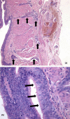
(a) Longitudinal slide, Hematoxylin and Eosin (HE) staining, 50x magnification: Abnormal glands (black arrows) in the submucosa reaching through the muscularis propria almost to the adventitia. (b) HE staining, 400x magnification: Pseudostratified columnar epithelium with cilia (black arrows).

References

    1. Nihoul-Fekete C, De Backer A, Lortat-Jacob S, Pellerin D. Congenital esophageal stenosis: A review of 20 cases. Pediatr Surg Int. 1987;2:86–92. doi: 10.1007/BF00174179. - DOI
    1. Kumar R. A case of congenital esophageal stricture due to a cartilaginous ring. Br J Surg. 1962;49:533–4. doi: 10.1002/bjs.18004921707. PMid: 14460500. - DOI - PubMed
    1. Anderson LS, Shackelford GD, Mancilla-Jimenez R, McAlister WH. Cartilaginous esophageal ring: a cause of esophageal stenosis in infants and children. Radiology. 1973;108(3):665–6. doi: 10.1148/108.3.665. PMid: 4198827. - DOI - PubMed
    1. Murphy SG, Yazbeck S, Russon P. Isolated congenital esophageal stenosis. J Pediatr Surg. 1995;30:1238–41. doi: 10.1016/0022-3468(95)90032-2. - DOI - PubMed
    1. Luedtke P, Levine MS, Rubesin SE, Weinstein DS, Laufer I. Radiologic diagnosis of benign esophageal strictures: a pattern approach. Radiographics. 2003;23(4):897–909. doi: 10.1148/rg.234025717. PMid: 12853664. - DOI - PubMed

LinkOut - more resources