Review
. 2018 Mar;80(1):11.21.1-11.21.17.
doi: 10.1002/cpph.36.

Three Decades of Chloride Intracellular Channel Proteins: From Organelle to Organ Physiology

Affiliations
Review

Three Decades of Chloride Intracellular Channel Proteins: From Organelle to Organ Physiology

Shubha Gururaja Rao et al. Curr Protoc Pharmacol. 2018 Mar.

Abstract

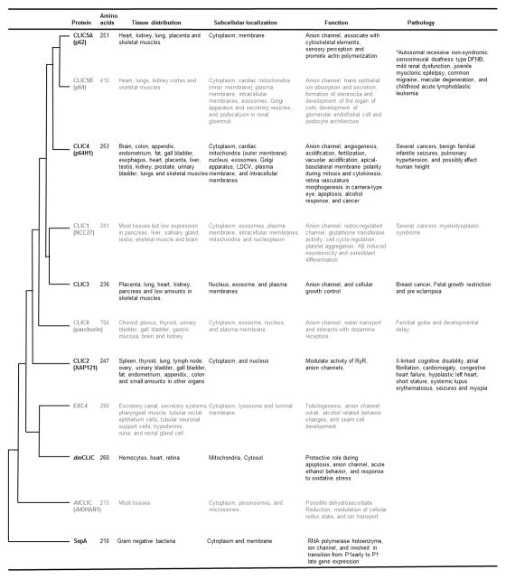
Intracellular organelles are membranous structures central for maintaining cellular physiology and the overall health of the cell. To maintain cellular function, intracellular organelles are required to tightly regulate their ionic homeostasis. Any imbalance in ionic concentrations can disrupt energy production (mitochondria), protein degradation (lysosomes), DNA replication (nucleus), or cellular signaling (endoplasmic reticulum). Ionic homeostasis is also important for volume regulation of intracellular organelles and is maintained by cation and anion channels as well as transporters. One of the major classes of ion channels predominantly localized to intracellular membranes is chloride intracellular channel proteins (CLICs). They are non-canonical ion channels with six homologs in mammals, existing as either soluble or integral membrane protein forms, with dual functions as enzymes and channels. Provided in this overview is a brief introduction to CLICs, and a summary of recent information on their localization, biophysical properties, and physiological roles. © 2018 by John Wiley & Sons, Inc.

Keywords: CLICs; chloride intracellular channels; intracellular organelles; ion channels; localization; mitochondria; physiology.


Figures

Figure 1
Figure 1. Summary of distribution, localization, physiological and pathological roles of family members of CLIC proteins
There are two major branches in the phylogenetic tree of mammalian CLICs, 1) comprising of CLIC2 which is not present in mice and 2) CLIC1, 3, 4, 5, and 6 present in all mammals. CLICs present in C. elegans and Drosophila are closely associated with mammalian CLICs. Bacterial and plant paralogs were recently characterized and are placed in distal branches of the phylogenetic tree. With the exception of CLIC6 and CLIC5B, all other CLIC paralogs are ~200–300 amino acids long. Tissue distribution of proteins is also indicated and CLICs are ubiquitous in expression. CLIC proteins are present in the cytoplasm as well as other intracellular organelles such as mitochondria, nucleus, exosomes and Golgi apparatus. At functional levels, CLICs form anion channels, participate in cellular physiology, and CLIC1 is known to have enzymatic activity. Changes in expression and roles of mammalian CLICs were demonstrated in several human pathological conditions such as cancer, epilepsy and congenital heart diseases.
Figure 2
Figure 2. Sequence and key features in the secondary structure of CLIC1
Key amino acids involved in ion channel properties and membrane anchoring are identified based on structure-function studies (Averaimo et al., 2013; Gururaja Rao et al., 2017; Hare, Goodchild, Breit, Curmi, & Brown, 2016; Liu et al., 2007; Singh & Ashley, 2006) and are highlighted with red and blue arrows within the transmembrane domain (grey box). The conserved nuclear localization signal (KKYR) is highlighted in the purple text. Grey dashed line indicates a salt bridge between R29 and E81 (Legg-E’silva et al., 2012). Key residues involved in modulation of channel activity are highlighted in red. Cholesterol Recognition/Interaction Amino Acid Consensus sequence (CRAC) are highlighted in the yellow box on the basis of specific published studies (Al Khamici et al., 2016) or predicted CRAC sequences (Fantini & Barrantes, 2013). Caveolin binding domain (black bar) (Byrne, Dart, & Rigden, 2012), PTMs (yellow, orange, pink, blue and green lines), 14-3-3 binding site (light blue box) (Johnson et al., 2010), and post-translational modification residues were identified using in silico analysis (Sigrist et al., 2010).

References

    1. Al-Momany A, Li L, Alexander RT, Ballermann BJ. Clustered PI(4,5)P(2) accumulation and ezrin phosphorylation in response to CLIC5A. J Cell Sci. 2014;127(Pt 24):5164–5178. doi: 10.1242/jcs.147744. - DOI - PubMed
    1. Al Khamici H, Brown LJ, Hossain KR, Hudson AL, Sinclair-Burton AA, Ng JP, … Valenzuela SM. Members of the chloride intracellular ion channel protein family demonstrate glutaredoxin-like enzymatic activity. PLoS One. 2015;10(1):e115699. doi: 10.1371/journal.pone.0115699. - DOI - PMC - PubMed
    1. Al Khamici H, Hossain KR, Cornell BA, Valenzuela SM. Investigating Sterol and Redox Regulation of the Ion Channel Activity of CLIC1 Using Tethered Bilayer Membranes. Membranes (Basel) 2016;6(4) doi: 10.3390/membranes6040051. - DOI - PMC - PubMed
    1. Andersen EF, Baldwin EE, Ellingwood S, Smith R, Lamb AN. Xq28 duplication overlapping the int22h-1/int22h-2 region and including RAB39B and CLIC2 in a family with intellectual and developmental disability. Am J Med Genet A. 2014;164A(7):1795–1801. doi: 10.1002/ajmg.a.36524. - DOI - PubMed
    1. Argenzio E, Margadant C, Leyton-Puig D, Janssen H, Jalink K, Sonnenberg A, Moolenaar WH. CLIC4 regulates cell adhesion and beta1 integrin trafficking. J Cell Sci. 2014;127(Pt 24):5189–5203. doi: 10.1242/jcs.150623. - DOI - PubMed

Publication types

Substances

LinkOut - more resources