. 2018;64(4):1307-1324.
doi: 10.3233/JAD-180514.

Diffuse Amyloid-β Plaques, Neurofibrillary Tangles, and the Impact of APOE in Elderly Persons' Brains Lacking Neuritic Amyloid Plaques

Affiliations

Diffuse Amyloid-β Plaques, Neurofibrillary Tangles, and the Impact of APOE in Elderly Persons' Brains Lacking Neuritic Amyloid Plaques

Erin L Abner et al. J Alzheimers Dis. 2018.

Abstract

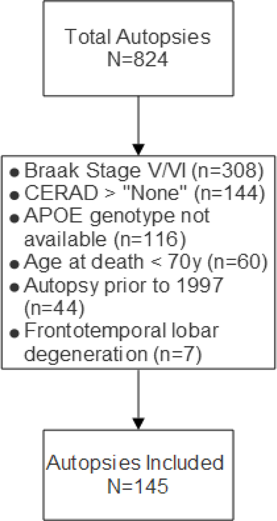
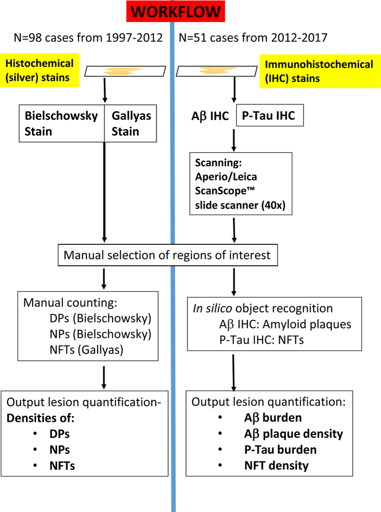
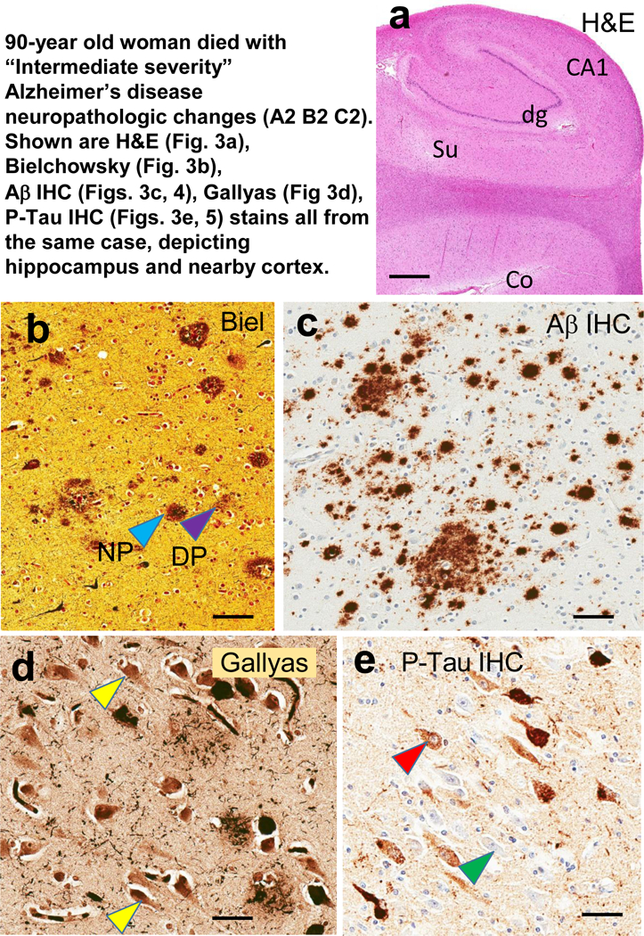
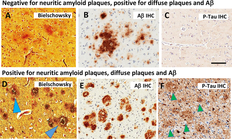
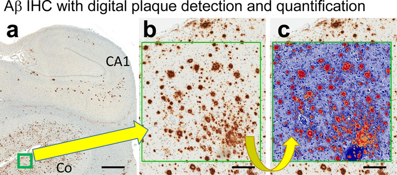
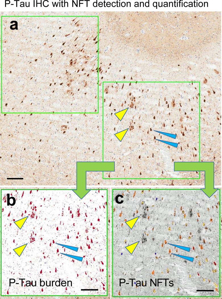
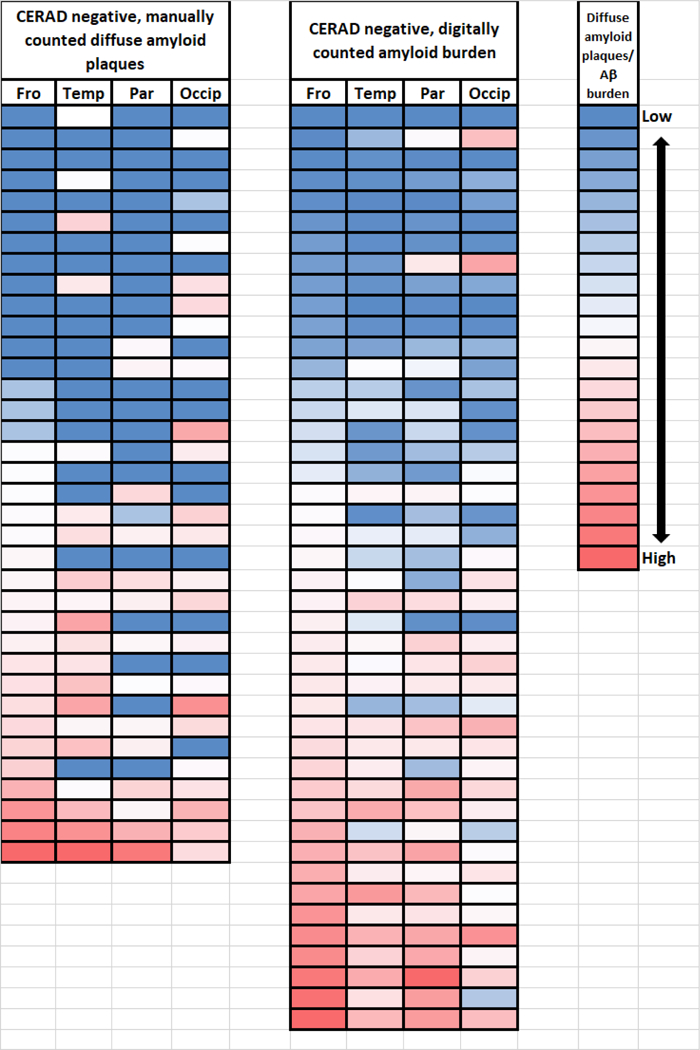
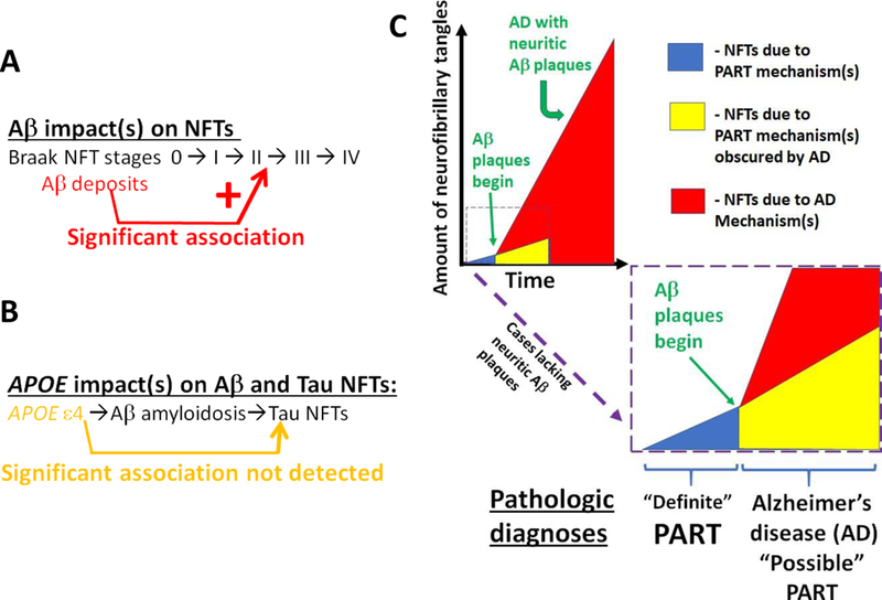
Data from a large autopsy series were analyzed to address questions pertinent to primary age-related tauopathy (PART) and Alzheimer's disease (AD): what factors are associated with increased severity of neurofibrillary degeneration in brains that lack neuritic amyloid plaques?; is there an association between Apolipoprotein E (APOE) alleles and PART pathologic severity independent of amyloid-β (Aβ) deposits?; and, how do the stains used to detect plaques and tangles impact the experimental results? Neuropathologic data were evaluated from elderly research volunteers whose brain autopsies were performed at University of Kentucky Alzheimer's Disease Center (UK-ADC; N = 145 subjects). All of the included subjects' brains lacked neuritic amyloid plaques according to the CERAD diagnostic criteria and the average final MMSE score before death was 26.8±4.6 stdev. The study incorporated evaluation of tissue with both silver histochemical stains and immunohistochemical stains to compare results; the immunohistochemical stains (Aβ and phospho-tau) were scanned and quantified using digital pathologic methods. Immunohistochemical stains provided important advantages over histochemical stains due to sensitivity and detectability via digital methods. When AD-type pathology was in its presumed earliest phases, neocortical parenchymal Aβ deposits were associated with increased medial temporal lobe neurofibrillary tangles. The observation supports the NIA-AA consensus recommendation for neuropathologic diagnoses, because even these "diffuse" Aβ deposits signal that AD pathobiologic mechanisms are occurring. Further, the data were most compatible with the hypothesis that the APOEɛ4 allele exerts its effect(s) via driving Aβ deposition, i.e., an "upstream" influence, rather than being associated directly with Aβ- independent PART pathology.

Keywords: Aging; Genie; MAPT; SNAP; ScanScope; amyloid-β; hippocampus; neuropathology; oldest-old.


Conflict of interest statement

Conflict of Interest/Disclosure Statement:

The authors have no conflict of interest to report

Figures

Fig. 1
Fig. 1. Flowchart of cases included, and bases for exclusions.
The University of Kentucky AD Center (UK-ADC) autopsy cohort is community-based and most patients were recruited while cognitively intact. Only 11% of the study participants were demented at final examination (See Table 1).
Fig. 2.
Fig. 2.. Workflow.
Schematic to depict the study design of the present study that included different UK-ADC cohorts and methods applied. Notably, different stains were used and the neuropathologic lesions were detected and counted separately. For the 1997–2012 cohort, histochemical stains were used with silver impregnation. Bielschowsky stains were used for diffuse amyloid plaques (DPs) and neuritic amyloid plaques (NPs), whereas the Gallyas silver impregnation technique was used for NFTs; for these cases, the output comprised manually tabulated density counts for DPs, NPs, and NFTs. For the 2012–2017 cohort, immunohistochemical (IHC) stains were used -- Aβ and P-Tau IHC – and slides scanned using a digital slide scanner, followed by in silico object recognition, with output including Aβ burden, Aβ plaque density, P-Tau burden, and P-Tau NFT density.
Fig. 3.
Fig. 3.. To illustrate characteristics of the different stains used, photomicrographs of stained sections from the hippocampus and nearby cortical structures are displayed.
These photomicrographs all depict brain sections from a woman who died at age 90 years with “Intermediate severity” Alzheimer’s disease neuropathologic changes (Thal Aβ stage 3, Braak NFT stage III, CERAD “moderate” density NPs). This case is only for illustrative purposes and was not included in the other analyses due to the presence of NPs. Adjacent sections from the same case were used for Figs. 3 and 4 also. On the hematoxylin and eosin (H&E)-stained section are indicated the structures in the tissue block corresponding to the hippocampal formation and adjacent structures: dentate granule cells (dg), CA1, subiculum (Su), and peri- allocortical cortex (Co). From the Co region is shown immunohistochemical stains used to characterize amyloid plaques: Bielschowsky silver stain (b) and Aβ IHC (c). Note in the Bielschowsky stain that one can discriminate NPs (pale blue arrowhead) from DPs (purple arrowhead), whereas this discrimination is not possible in the Aβ IHC (c). Nonetheless, note that there is increased sensitivity and enhanced signal:noise ratio in the Aβ IHC stain compared to silver stains. For staining NFTs in CA1, the Gallyas silver stain (d) and P-Tau IHC (e) were used. Note that the signal: noise ratio is not high for the Gallyas stain (d); some of the structures may or may not represent NFTs (yellow arrowheads). This signal:noise ratio confounds digital assessment. By contrast, in the P-Tau IHC (e), even the pre-tangles (red arrowhead) are far easier to discriminate than the non-affected neurons (green arrowhead). Scale bars = 3mm (a); = 100 microns (b,c); and, = 50 microns (d,e).
Fig. 4.
Fig. 4.. Depiction of stains in a case that lack neuritic amyloid plaques (Panels A-C) in comparison to a case with neuritic amyloid plaques (D-F).
The Bielschowsky silver stain labels amorphous, fleecy material in the brain of an 80 year old woman; immunohistochemistry confirms the presence of Aβ (B; Aβ IHC), whereas P-Tau IHC is negative for dystrophic neurites. In AD, by contrast, the Bielschowsky stain highlights plaques invested with degenerating neurites (pale blue arrowhead) and/or ringing a discrete core (dark blue arrowhead). Note that Aβ IHC is immunopositive (E), and the P-Tau IHC in this case highlights the profiles of neuritic amyloid plaques (green arrowheads in panel F).
Fig. 5.
Fig. 5.. Photomicrographs depict an area used to evaluate Aβ IHC using digital pathology.
Low-power photomicrograph of Aβ IHC stained section (a) shows where the boxed area of interest was chosen. At higher magnification (b), the Aβ immunostained structures can be easily observed. The digitally false-colored box (c) shows how the Aβ plaques are digitally recognized; all of the orange, red, and yellow areas are quantified automatically. Scale bars =3mm (a); = 150 microns (b,c).
Fig. 6.
Fig. 6.. Photomicrographs depict an area used to evaluate P-Tau IHC using digital pathology.
Low-power photomicrograph of P-Tau IHC stained section (a) depicts the size of the boxes (two such boxes are shown here, up to ten are used per slide) that are used to indicate regions of interest for the digital quantificaiton. At higher power (b-c), digital renderings of the lower-right box in panel (a) are presented to show the areas recognized for P-Tau burden (red in panel b); and P-Tau NFTs (orange in panel c). The yellow arrowheads in all 3 panels show the same NPs, and blue arrowheads show two specific NFTs. Note that the P-Tau burden image recognition algorithm (b) incorporated the NP structures, whereas the P-Tau NFTs highlights only the NFTs, not the NPs. Scale bars = 200 microns (a-c).
Fig. 7.
Fig. 7.. Quantitative pathology in four different neocortical brain regions for cases that were CERAD-negative for neuritic amyloid plaques.
Shown are color-coded (red=high; white=middle; blue=low) representations of the cases counted manually as diffuse amyloid plaques (1997–2012; n=36) and digitally quantified Aβ amyloid burden (2012–2017; n=44), depicting the severity and distribution of those pathologies in the frontal (Fro; Brodmann areas 39/40); superior and middle temporal (Temp; Brodmann areas 2½2); parietal (Par; Brodmann area xx); and, occipital (Occip; Brodmann areas 17/18) neocortical regions. Each row represents one brain. The cases are ranked from top to bottom for both cohorts in the order from lowest to highest amount of detected pathology in the frontal cortical sections.
Fig. 8.
Fig. 8.. Overview of the results.
The data and analyses provide support for the hypothesis that Aβ deposits, even in the earliest “diffuse” phase, are associated with increased severity of medial temporal lobe NFT pathology as assessed by digital pathology of immunohistochemical stains (A). By contrast, when Aβ pathology was taken into account, there was not evidence for direct and robust association between APOE genotype and the severity of tau/NFT pathology (B). An over-arching point, as depicted in schematic form (C), is that in cases with early AD, both AD- related and PART-related pathogenetic mechanisms are probably occurring at the same time. Whereas it may be most specific to use Aβ immunohistochemistry for Definite PART, our analyses indicate it still is accurate to assume that some PART pathology is probably present despite the presence of Aβ plaques.

References

    1. Nelson PT, Jicha GA, Schmitt FA, Liu H, Davis DG, Mendiondo MS, Abner EL, Markesbery WR (2007) Clinicopathologic correlations in a large Alzheimer disease center autopsy cohort: neuritic plaques and neurofibrillary tangles “do count” when staging disease severity. J Neuropathol Exp Neurol 66, 1136–1146. - PMC - PubMed
    1. Kovacs GG, Milenkovic I, Wohrer A, Hoftberger R, Gelpi E, Haberler C, Honigschnabl S, Reiner-Concin A, Heinzl H, Jungwirth S, Krampla W, Fischer P, Budka H (2013) Non-Alzheimer neurodegenerative pathologies and their combinations are more frequent than commonly believed in the elderly brain: a community-based autopsy series. Acta Neuropathol 126, 365–384. - PubMed
    1. Crary JF, Trojanowski JQ, Schneider JA, Abisambra JF, Abner EL, Alafuzoff I, Arnold SE, Attems J, Beach TG, Bigio EH, Cairns NJ, Dickson DW, Gearing M, Grinberg LT, Hof PR, Hyman BT, Jellinger K, Jicha GA, Kovacs GG, Knopman DS, Kofler J, Kukull WA, Mackenzie IR, Masliah E, McKee A, Montine TJ, Murray ME, Neltner JH, Santa-Maria I, Seeley WW, Serrano-Pozo A, Shelanski ML, Stein T, Takao M, Thal DR, Toledo JB, Troncoso JC, Vonsattel JP, White CL 3rd, Wisniewski T, Woltjer RL, Yamada M, Nelson PT(2014) Primary age-related tauopathy (PART): a common pathology associated with human aging. Acta Neuropathol 128, 755–766. - PMC - PubMed
    1. Bouras C, Hof PR, Morrison JH (1993) Neurofibrillary tangle densities in the hippocampal formation in a non-demented population define subgroups of patients with differential early pathologic changes. Neurosci Lett 153, 131–135. - PubMed
    1. Arriagada PV, Marzloff K, Hyman BT (1992) Distribution of Alzheimer-type pathologic changes in nondemented elderly individuals matches the pattern in Alzheimer’s disease. Neurology 42, 1681–1688. - PubMed

Publication types

MeSH terms