. 2018 Oct:73:21-33.
doi: 10.1016/j.bbi.2018.07.017. Epub 2018 Jul 21.

The concerted amyloid-beta clearance of LRP1 and ABCB1/P-gp across the blood-brain barrier is linked by PICALM

Affiliations

The concerted amyloid-beta clearance of LRP1 and ABCB1/P-gp across the blood-brain barrier is linked by PICALM

Steffen E Storck et al. Brain Behav Immun. 2018 Oct.

Abstract

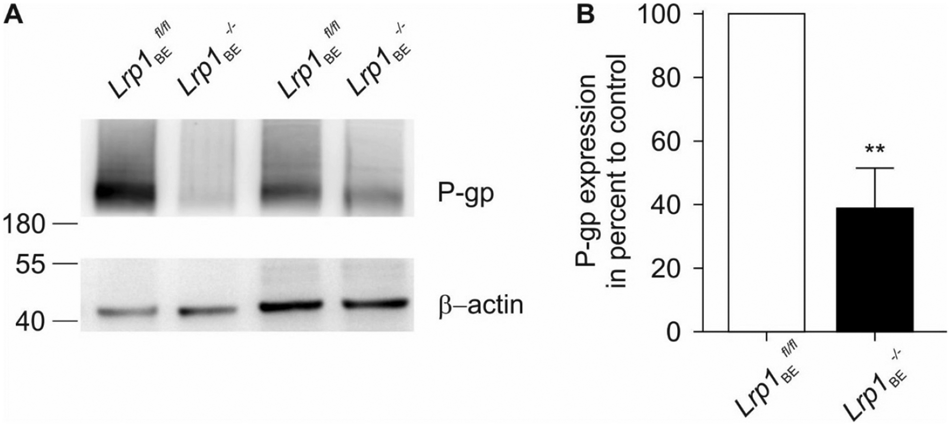
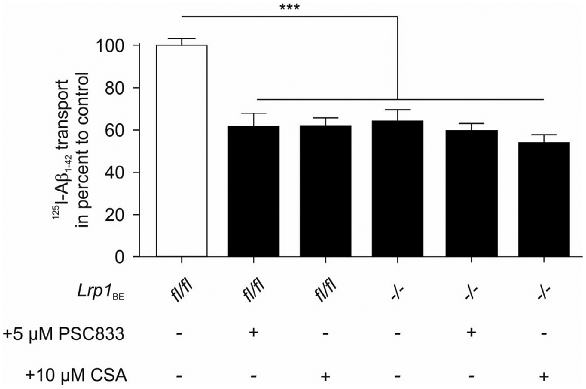
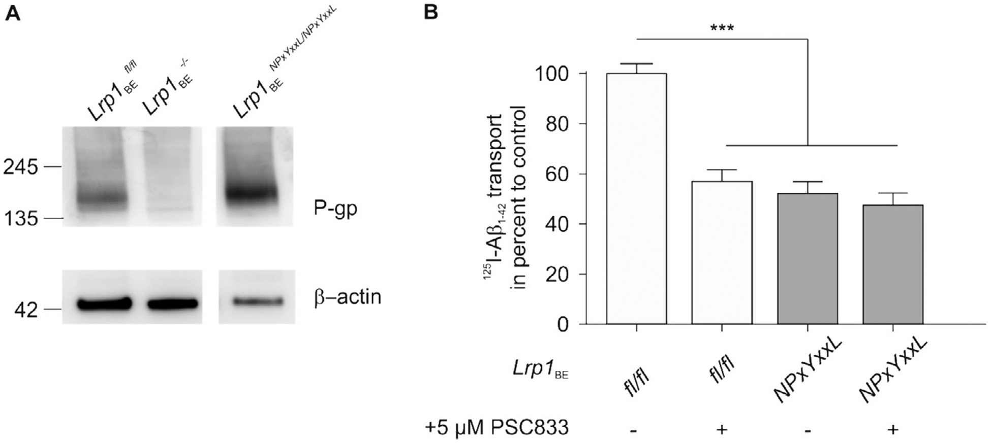
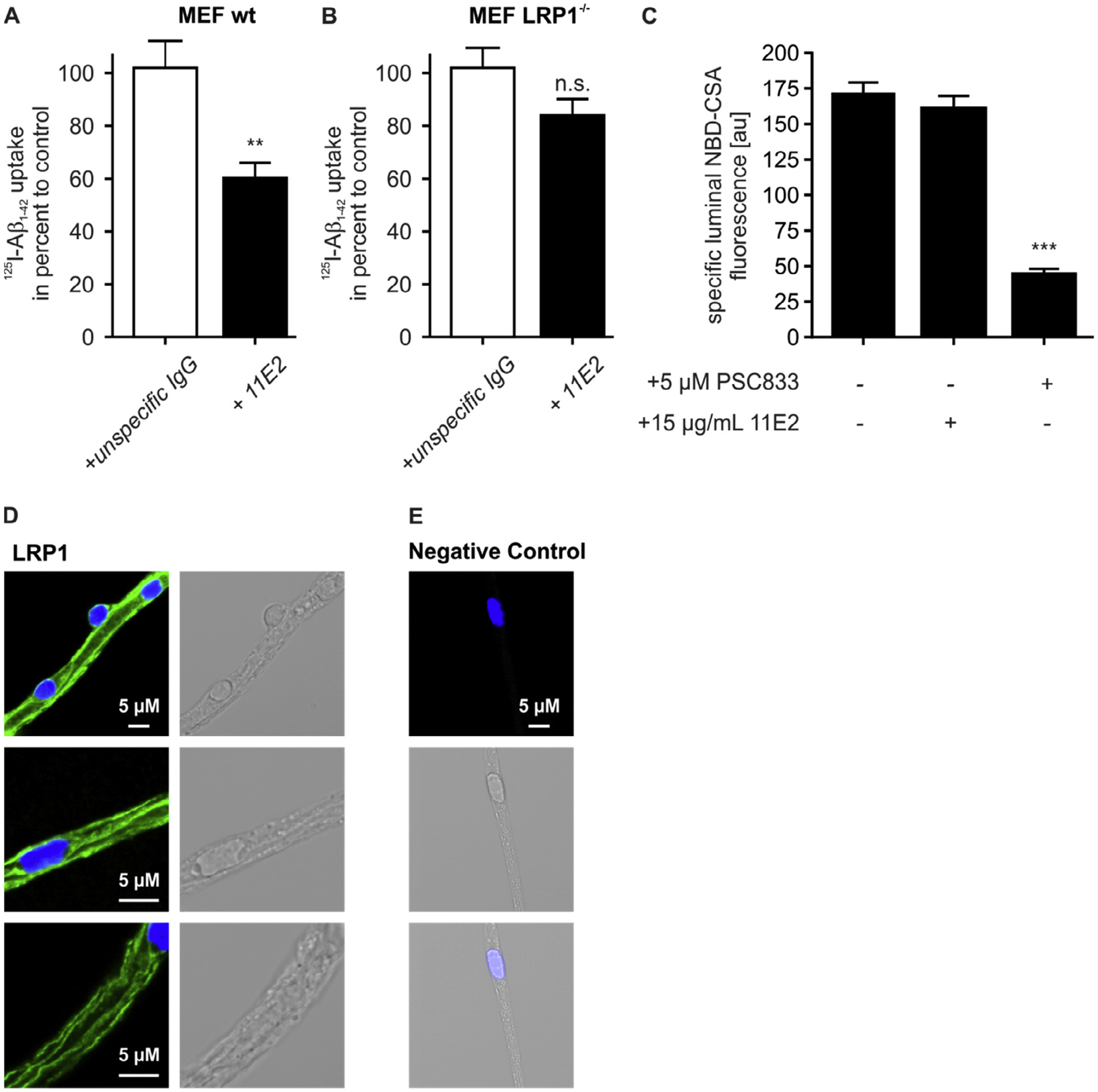
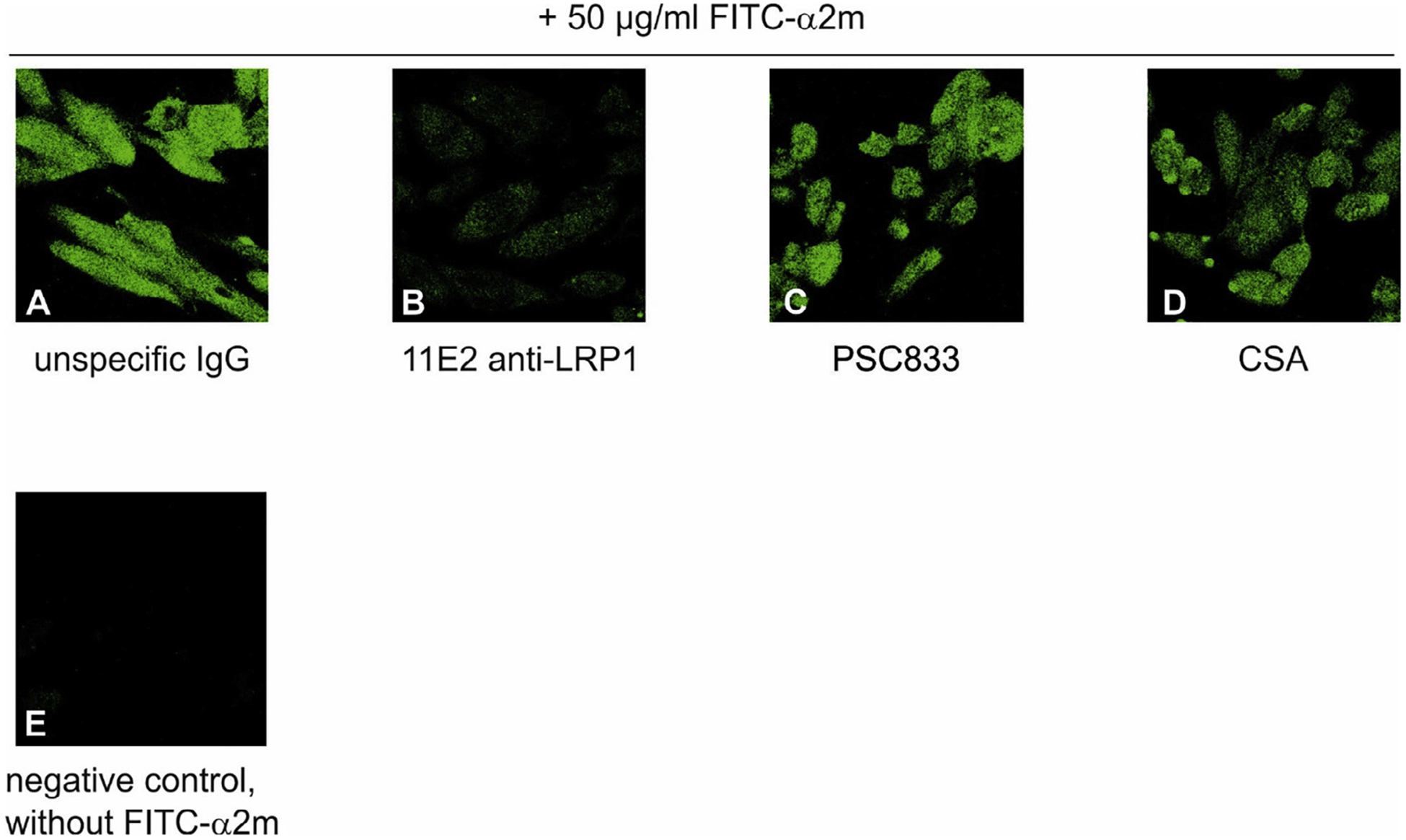
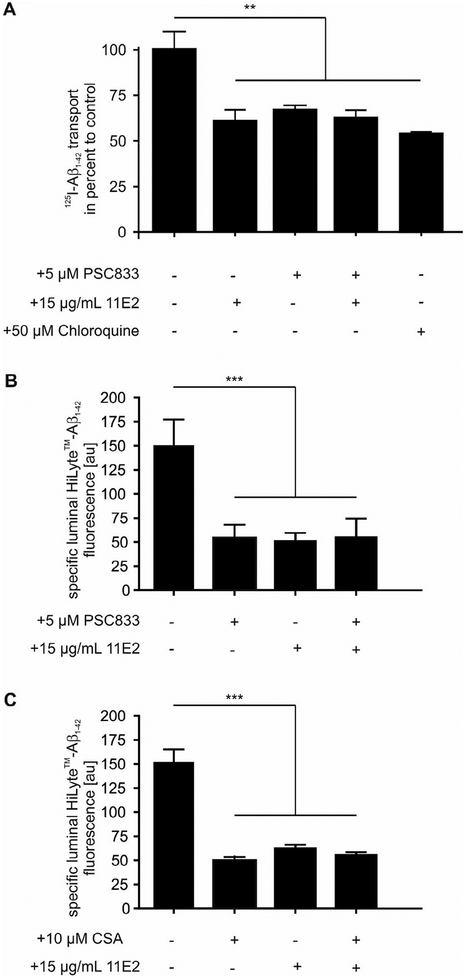
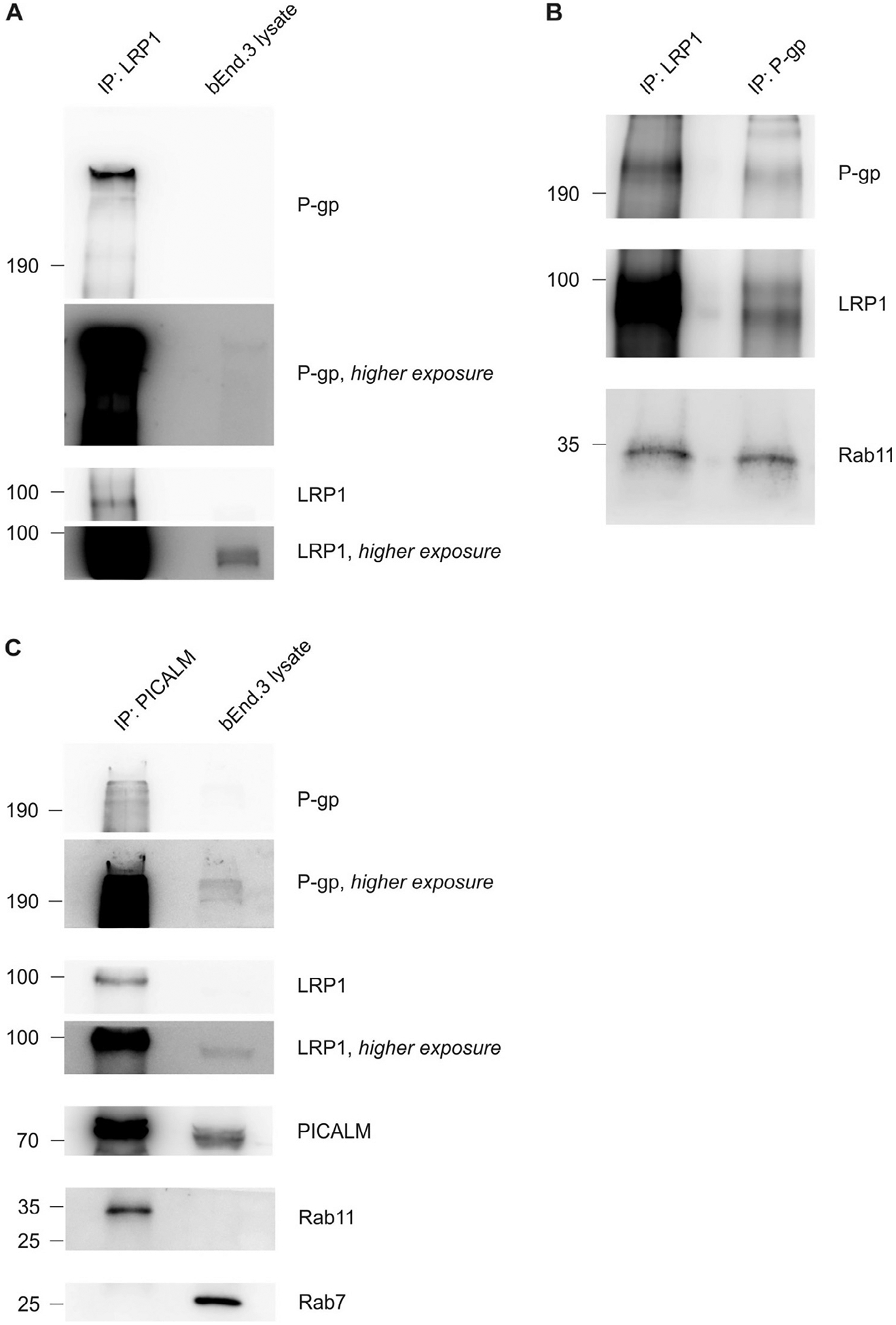
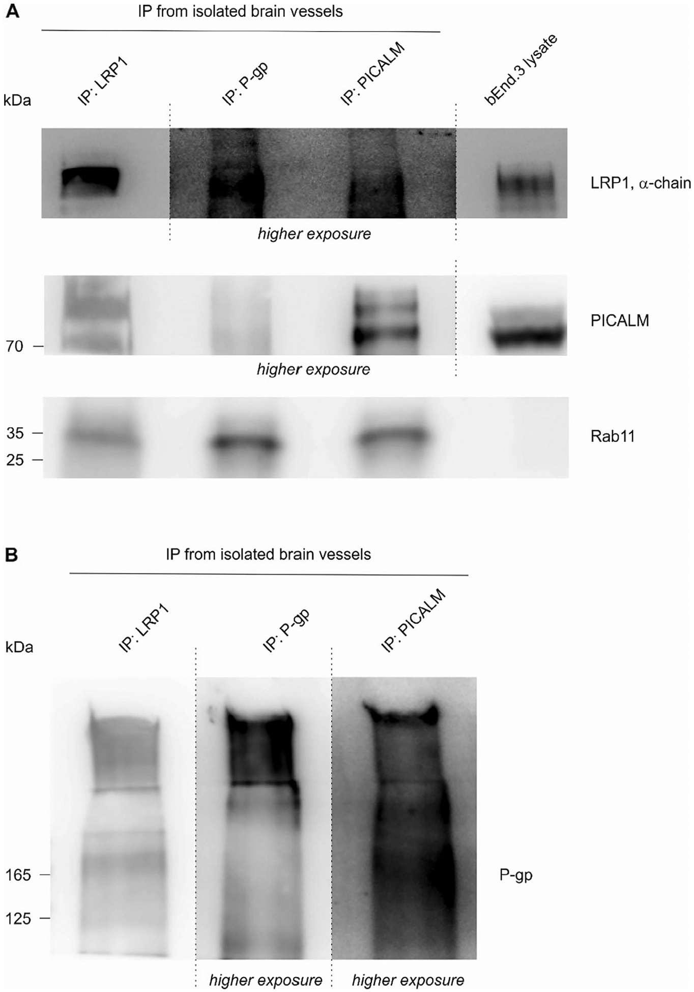
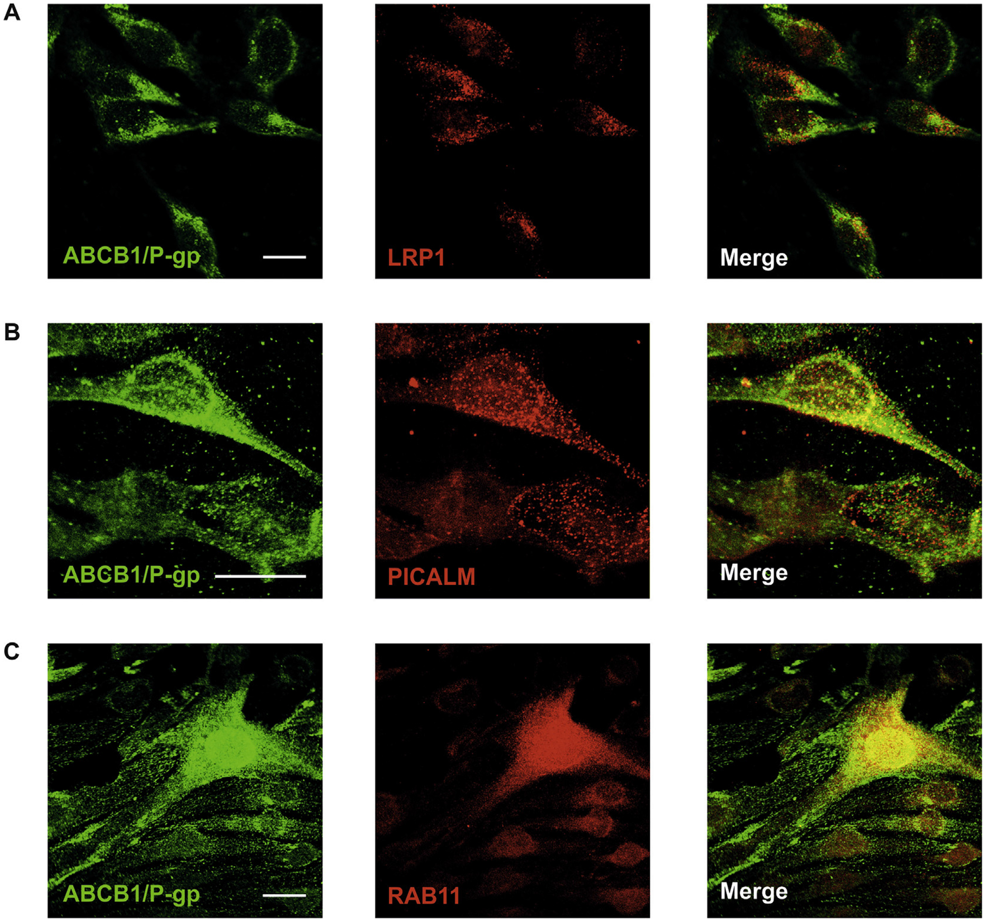
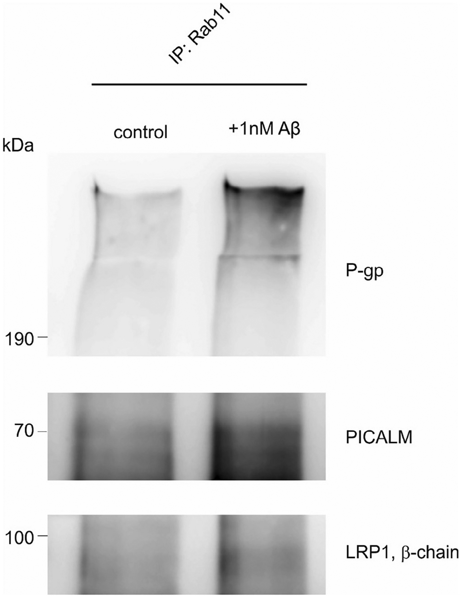
The accumulation of neurotoxic amyloid-beta (Aβ) in the brain is a characteristic hallmark of Alzheimer's disease (AD). The blood-brain barrier (BBB) provides a large surface area and has been shown to be an important mediator for removal of brain Aβ. Both, the ABC transporter P-glycoprotein (ABCB1/P-gp) and the receptor low-density lipoprotein receptor-related protein 1 (LRP1) have been implicated to play crucial roles in Aβ efflux from brain. Here, with immunoprecipitation experiments, co-immunostainings and dual inhibition of ABCB1/P-gp and LRP1, we show that both proteins are functionally linked, mediating a concerted transcytosis of Aβ through endothelial cells. Late-onset AD risk factor Phosphatidylinositol binding clathrin assembly protein (PICALM) is associated with both ABCB1/P-gp and LRP1 representing a functional link and guiding both proteins through the brain endothelium. Together, our results give more mechanistic insight on Aβ transport across the BBB and show that the functional interplay of different clearance proteins is needed for the rapid removal of Aβ from the brain.

Keywords: ABC transporter B1/P-glycoprotein (ABCB1/P-gp); Alzheimer’s disease (AD); Amyloid-beta (Aβ); Blood-brain barrier (BBB); Clearance; Endothelial cell; Endothelium; Low-density lipoprotein receptor-related protein 1 (LRP1); Phosphatidylinositol-binding clathrin assembly protein (PICALM); Transcytosis.


Figures

Fig. 1.
Fig. 1.
ABCB1/P-gp expression is decreased in Lrp1 knockout capillaries. (A) Representative immunoblots of ABCB1/P-gp in ex vivo capillaries isolated from control (Lrp1BEfl/fl) and brain endothelial-specific Lrp1 knockout (Lrp1BE−/−) mice. An anti-β-actin immunoblot is shown as a loading control. (B) Quantification of relative abundance of ABCB1/P-gp expression of n = 5 independent experiments. For each isolation ≥3 mice per group were used (mean ± SEM). For statistical analyses, unpaired t test was used. **P < 0.01.
Fig. 2.
Fig. 2.
No effect of ABCB1/P-gp inhibition on [125I] Aβ1−42 transcytosis across Lrp1 knockout endothelial cells. [125I]-Aβ1−42 transport across LRP1 knockout (Lrp1BE−/−) and control (Lrp1BEfl/fl) primary mouse brain capillary endothelial cell monolayer was studied in the presence of 1 μCi/ml [14C]-inulin to normalize for passive diffusion. Transcytosis was analyzed in the brain-to-blood direction (abluminal to luminal) by measuring the dpm for [14C]-inulin and the cpm for the TCA-precipitable [125I]-radioactivity. Transport rates were normalized to transport rates of control Lrp1BEfl/fl brain endothelial cells in the absence of any inhibitors. Transport was studied at a physiological concentration of 0.1 nM [125I]-Aβ1−42 and 5 μM PSC833 or 10 μM Cyclosporine A (CSA). Data represent mean ± SEM of n = 18; n = 21, n = n = 27, n = 17, n = 11 from left to right of at least 2 independent experiments. For statistical analyses, repeated-measures ANOVA followed by Bonferroni multiple comparisons was used. ***P < 0.001.
Fig. 3.
Fig. 3.
Despite enhanced ABCB1/P-gp expression in LRP1 NPxYxxL knock-in endothelial cells, ABCB1/P-gp inhibition has no effect on [125I] Aβ1−42 transcytosis. (A) Immunoblot analysis of ABCB1/P-gp in ex vivo capillaries isolated from control (Lrp1BEfl/fl), endothelial-specific Lrp1 knockout (Lrp1BE−/−) and LRP1 NPxYxxL knock-in (Lrp1BENPxYxxL/NPxYxxL) mice. Representative result of n = 2 independent isolations with n ≥ 3 mice per group. Lysates were analyzed on the same Western blot but rearranged for clearer presentation(B) [125I]-Aβ1−42 transport across the primary mouse brain capillary endothelial cell monolayer was studied in the presence of 1 μCi/ml [14C]-inulin to normalize for passive diffusion. Transcytosis was analyzed in the brain-to-blood direction (abluminal to luminal) by measuring the dpm for [14C]-inulin and the cpm for the TCA-precipitable [125I] radioactivity. Transport rates were normalized to transport rates of Lrp1BEfl/fl brain endothelial cells in the absence of PSC833. Transport was studied at a physiological concentration of 0.1 nM [125I]-Aβ1−42. Data represent mean ± SEM of n = 12; n = 20, n = 19, n = 14 from left to right of 3 independent experiments. For statistical analyses, repeated-measures ANOVA followed by Bonferroni multiple comparisons was used. ***P < 0.001.
Fig. 4.
Fig. 4.
11E2 anti-LRP1 antibody is a potent LRP1 inhibitor that does not interfere with ABCB1/P-gp function. (A) 11E2 anti-LRP1 antibody reduces the uptake of 0.1 nM [125I] Aβ1−42 in wt mouse embryonic fobroblasts (MEF wt) but (B) has no effect on the uptake of 0.1 nM [125I] Aβ1−42 in Lrp1-defecient mouse embryonic fobroblasts (MEF LRP1/) results of (A) and (B) represent the mean and SEM of n ≥ 9 of one experiment. (C) 11E2 anti-LRP1 antibody has no effect on ABCB1/P-gp function. For luminal NBD-CSA fluorescence, each data point represents the mean value of 7 capillaries from a single preparation (pooled tissue from 30 CD-1 mice); variability is given by SEM bars. Units are arbitrary fluorescence units (scale 0–255). (D) Immunofluorescent staining for LRP1 in isolated brain capillaries. Scale bar: 5 μm. For statistical analyses, repeated-measures ANOVA followed by Bonferroni multiple comparisons was used. **P < 0.01, ***P < 0.001.
Fig. 5.
Fig. 5.
ABCB1/P-gp inhibitors PSC833 and CSA do not affect LRP1 function Uptake of FITC-labeled alpha-macroglobulin (FITC-α2m) into LRP1-expressing CHO K1 cells. Cells were incubated in the presence of 50 μg/ml FITC-α2m and 15 μg/mL unspecific IgG (A), 15 μg/mL 11E2 anti-LRP1 (B), 5 μM PSC833 (C) or 10 μM Cyclosporine A (CSA) for 60 min, washed, fixed and subjected to fluorescence detection. Negative control without FITC-α2m (D).
Fig. 6.
Fig. 6.
Simultaneous inhibition of LRP1 and ABCB1/P-gp has no additional effect on Aβ1−42 transcytosis. (A) Aβ1−42 transport across a primary porcine brain endothelial monolayer [125I]-Aβ1−42 transport was studied in the presence of 1 μCi/ml [14C]-inulin to normalize for passive diffusion. Transcytosis was analyzed in the brain-to-blood direction (abluminal to luminal) by measuring the dpm for [14C]-inulin and the cpm for the TCA-precipitable [125I] radioactivity. Transport rates were normalized to transport rates of untreated brain endothelial cells. Transport was studied at a physiological concentration of 0.2 nM [125I]-Aβ1−42. For ABCB1/P-gp Inhibition 5 μM PSC833 and for LRP1 inhibition 20 μg/mL 11E2 anti-LRP1 was used. Data represent mean ± SEM of n = 12; n = 20, n = 19, n = 14 from left to right of two independent experiment. The chloroquine group represents data of n = 4 of one experiment. (B + C) Aβ1−42 transport in isolated capillaries. For luminal HiLyte-Aβ1–42 fluorescence, each data point represents the mean value of 10 capillaries from a single preparation (pooled tissue from 30 CD-1 mice); variability is given by SEM bars. For ABCB1/P-gp Inhibition 5 μM PSC833 (B) or 10 μM Cyclosporine A (CSA) (C) and for LRP1 inhibition 15 μg/mL 11E2 anti-LRP1 was used. Units are arbitrary fluorescence units (scale 0–255). For statistical analyses, repeated-measures ANOVA followed by Bonferroni multiple comparisons was used. **P < 0.01; ***P < 0.001.
Fig. 7.
Fig. 7.
Co-immunoprecipitation of LRP1, ABCB1/P-gp and PICALM. (A and B) Co-immunoprecipitation of LRP1 and ABCB1/P-gp in brain endothelial cell line bEnd.3 using different antibodies. Rab11 can be co-immunoprecipitated with both ABCB1/P-gp and LRP1. (C) Co-immunoprecipitation of PICALM and LRP1, ABCB1/P-gp, Rab11 but not Rab7. Representative results of experiments done twice (A + B), Data shown from the ABCB1/P-gp-IP (B) is the result of one experiment.
Fig. 8.
Fig. 8.
Co-immunoprecipitation of LRP1, ABCB1/P-gp, PICALM and Rab11 from isolated capillaries. (A + B) The three proteins LRP1, ABCB1/P-gp are associated with each other and are present in Rab11-positive vesicles in isolated capillaries, Data shown is the result of one experiment from capillaries isolated from 10 mice.
Fig. 9.
Fig. 9.
Colocalization of ABCB1/P-gp with other proteins involved in Aβ transport. ABCB1/P-gp co-localizes with LRP1 (A), PICALM (B) and Rab11 (C) in bEnd.3 cells. Scale bar: 20 μm.
Fig. 10.
Fig. 10.
Co-immunoprecipitation of LRP1, ABCB1/P-gp and PICALM with Rab11 after Aβ stimulation. bEnd3 were stimulated with 1 nM Aβ1–40 for 5 min.

References

    1. Aleksis R, Oleskovs F, Jaudzems K, Pahnke J, Biverstal H, 2017. Structural studies of amyloid-beta peptides: unlocking the mechanism of aggregation and the associated toxicity. Biochimie 140, 176–192. - PubMed
    1. Bateman RJ, Munsell LY, Morris JC, Swarm R, Yarasheski KE, Holtzman DM, 2006. Human amyloid-beta synthesis and clearance rates as measured in cerebrospinal fluid in vivo. Nat. Med 12 (7), 856–861. - PMC - PubMed
    1. Bell RD, Deane R, Chow N, Long X, Sagare A, Singh I, Streb JW, Guo H, Rubio A, Van Nostrand W, Miano JM, Zlokovic BV, 2009. SRF and myocardin regulate LRP-mediated amyloid-beta clearance in brain vascular cells. Nat. Cell Biol 11 (2), 143–153. - PMC - PubMed
    1. Bell RD, Sagare AP, Friedman AE, Bedi GS, Holtzman DM, Deane R, Zlokovic BV, 2007. Transport pathways for clearance of human Alzheimer’s amyloid beta-peptide and apolipoproteins E and J in the mouse central nervous system. J. Cereb. Blood Flow Metab 27 (5), 909–918. - PMC - PubMed
    1. Cirrito JR, Deane R, Fagan AM, Spinner ML, Parsadanian M, Finn MB, Jiang H, Prior JL, Sagare A, Bales KR, Paul SM, Zlokovic BV, Piwnica-Worms D, Holtzman DM, 2005. P-glycoprotein deficiency at the blood-brain barrier increases amyloid-beta deposition in an Alzheimer disease mouse model. J. Clin. Invest 115 (11), 3285–3290. - PMC - PubMed

Publication types

MeSH terms

Substances