

Near-Infrared Fluorescent Proteins: Multiplexing and Optogenetics across Scales
- PMID: 30041828
- PMCID: PMC6240479
- DOI: 10.1016/j.tibtech.2018.06.011
Near-Infrared Fluorescent Proteins: Multiplexing and Optogenetics across Scales
Abstract
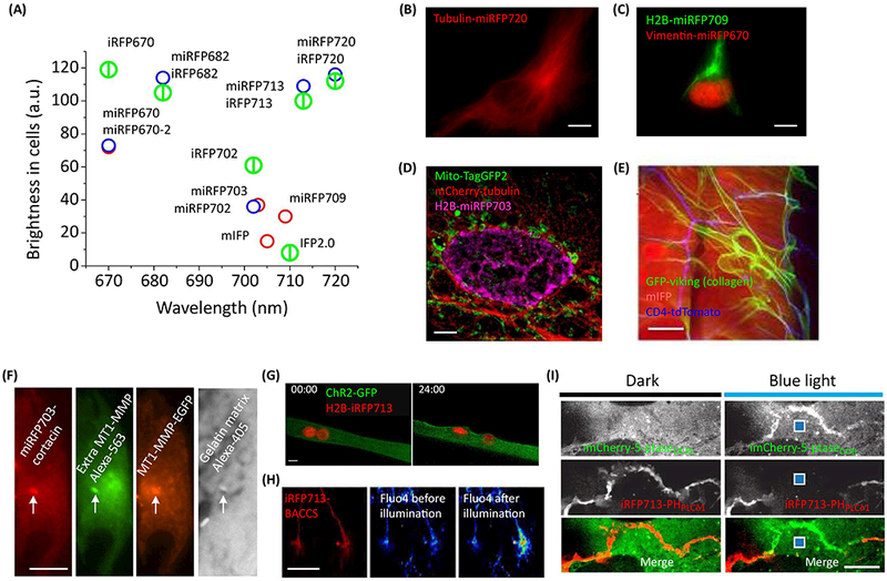
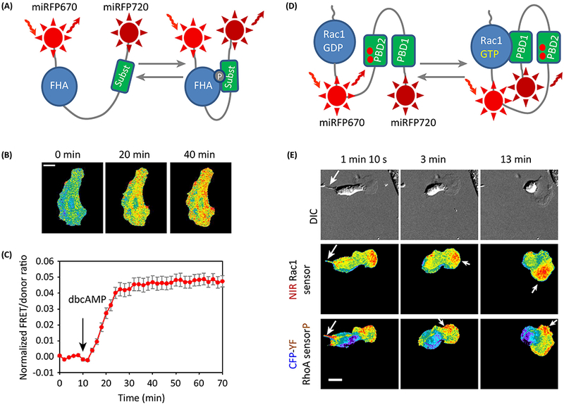
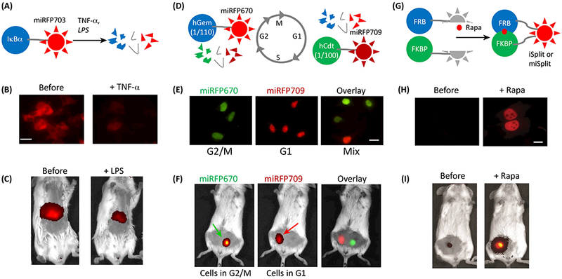
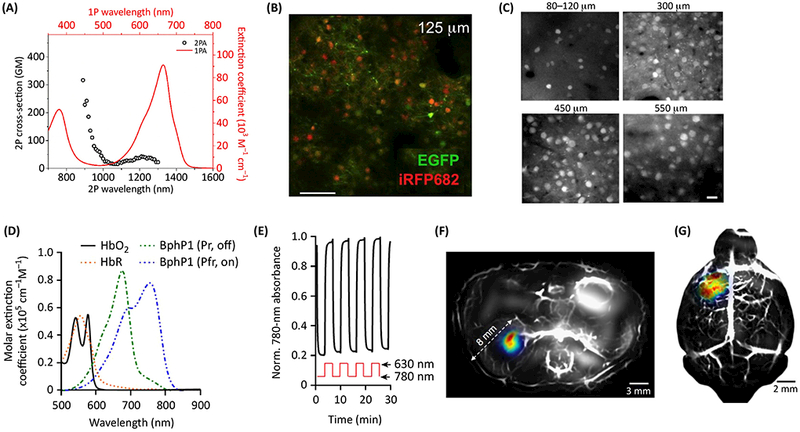
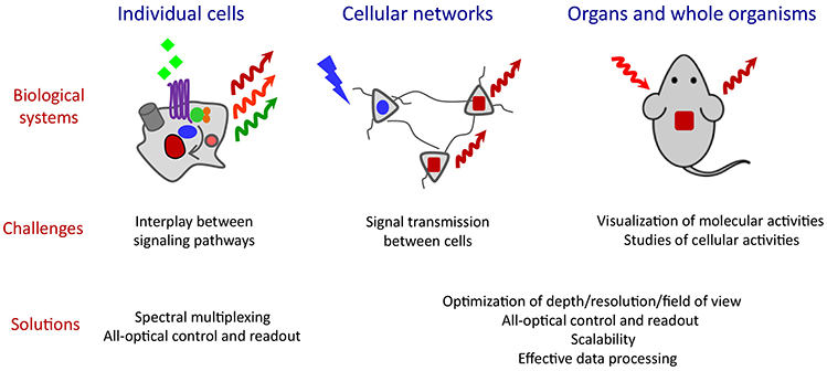
Since mammalian tissue is relatively transparent to near-infrared (NIR) light, NIR fluorescent proteins (FPs) engineered from bacterial phytochromes have become widely used probes for non-invasive in vivo imaging. Recently, these genetically encoded NIR probes have been substantially improved, enabling imaging experiments that were not possible previously. Here, we discuss the use of monomeric NIR FPs and NIR biosensors for multiplexed imaging with common visible GFP-based probes and blue light-activatable optogenetic tools. These NIR probes are suitable for visualization of functional activities from molecular to organismal levels. In combination with advanced imaging techniques, such as two-photon microscopy with adaptive optics, photoacoustic tomography and its recent modification reversibly switchable photoacoustic computed tomography, NIR probes allow subcellular resolution at millimeter depths.
Keywords: all-optical electrophysiology; bacterial phytochrome; biosensor; deep-tissue imaging; iRFP.
Copyright © 2018 Elsevier Ltd. All rights reserved.
Conflict of interest statement
Competing financial interests
Figures
References
-
- Jiguet-Jiglaire C et al. (2014) Noninvasive near-infrared fluorescent protein-based imaging of tumor progression and metastases in deep organs and intraosseous tissues. J Biomed Opt 19 (1), 16019. - PubMed
-
- Lai CW et al. (2016) Using Dual Fluorescence Reporting Genes to Establish an In Vivo Imaging Model of Orthotopic Lung Adenocarcinoma in Mice. Mol Imaging Biol 18 (6), 849–859. - PubMed
Publication types
MeSH terms
Substances
Grants and funding
LinkOut - more resources
Full Text Sources
Other Literature Sources
Miscellaneous
