Review
. 2018 Sep;22(9):752-763.
doi: 10.1016/j.tics.2018.06.009. Epub 2018 Jul 23.

Social Origins of Cortical Face Areas

Affiliations
Review

Social Origins of Cortical Face Areas

Lindsey J Powell et al. Trends Cogn Sci. 2018 Sep.

Abstract

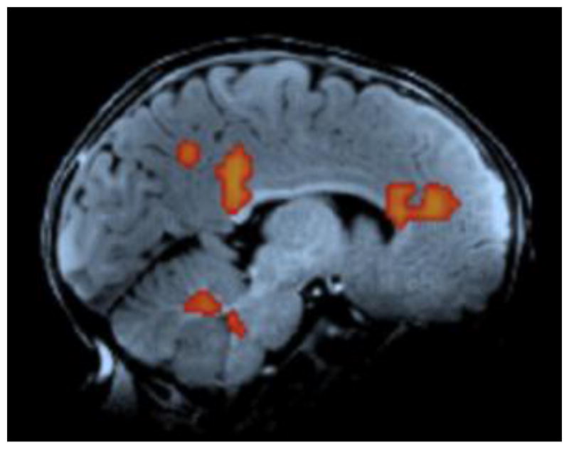
Recently acquired fMRI data from human and macaque infants provide novel insights into the origins of cortical networks specialized for perceiving faces. Data from both species converge: cortical regions responding preferentially to faces are present and spatially organized early in infancy, although fully selective face areas emerge much later. What explains the earliest cortical responses to faces? We review two proposed mechanisms: proto-organization for simple shapes in visual cortex, and an innate subcortical schematic face template. In addition, we propose a third mechanism: infants choose to look at faces to engage in positively valenced, contingent social interactions. Activity in medial prefrontal cortex during social interactions may, directly or indirectly, guide the organization of cortical face areas.

Keywords: cognitive neuroscience; development; faces; infancy; social interaction.


Figures

Figure I (Box 2)
Figure I (Box 2)
MPFC response to dynamic faces in human infants.
Figure 1
Figure 1. Face preferring regions across human development
Functional MRI reveals a similar location for regions that preferentially respond to faces (hot colors) when contrasted with scenes (cool colors), across development (top row). Selective responses to faces (hot colors) when contrasted with objects (cool colors) emerge later in development with size and selectivity of these regions increasing through adolescence (bottom row). Peak clusters are displayed on exemplar subjects for illustrative purposes. Data from 5- and 10-year-old children were provided by Michael Cohen and Nancy Kanwisher; infant and adult data were reported in [8].
Figure 2
Figure 2. Three mechanisms guiding development of cortical face areas
a) Extrastriate cortex is organized according to overlapping low-level feature dimensions (e.g. the eccentricity and shape dimensions represented orthogonally along the x and y axes) [33]. Preferential responses to faces may arise in the part of extrastriate cortex ("Face Area", outlined) that combines their particular low-level properties (e.g. frequent foveation and curvilinearity). b) An innate subcortical template may facilitate detection of face-like images. The template could influence face areas either by directing infants' visual attention, or through direct connectivity (shown here). Biased connectivity from this template could potentiate responses to faces in specific regions of extrastriate cortex (i.e. the “Face Area” box in this panel). c) Infants' preferential interest in social interaction, and the brain systems that support such interactions, may also influence face area development either by directing infants' visual attention, or through direct connectivity (shown here). In particular, biased connectivity from medial prefrontal cortex (MPFC) regions that respond to prosocial interaction could potentiate responses to faces in specific regions of extrastriate cortex (i.e. the “Face Area” box in this panel).

Comment in

References

    1. Kanwisher N, et al. The Fusiform Face Area: A Module in Human Extrastriate Cortex Specialized for Face Perception. J Neurosci. 1997;17:4302–4311. - PMC - PubMed
    1. Mccarthy G, et al. Face-Specific Processing in the Human Fusiforrn Gyms. J Cogn Neurosci. 1997;9:605–610. - PubMed
    1. Kanwisher N. The Quest for the FFA and Where It Led. J Neurosci. 2017;37:1056–1061. - PMC - PubMed
    1. Tsao DY, et al. Faces and objects in macaque cerebral cortex. Nat Neurosci. 2003;6:989–995. - PMC - PubMed
    1. Tsao DY, et al. Comparing face patch systems in macaques and humans. Proc Natl Acad Sci. 2008;105:19514–19519. - PMC - PubMed

Publication types

LinkOut - more resources