

doi: 10.1038/s41375-018-0220-z.
Epub 2018 Jul 24.
Identification of immune-activated hematopoietic stem cells
Affiliations
- PMID: 30042413
- PMCID: PMC6127088
- DOI: 10.1038/s41375-018-0220-z
Item in Clipboard
Identification of immune-activated hematopoietic stem cells
Leukemia.
2018 Sep.
No abstract available
Conflict of interest statement
The authors declare that they have no conflict of interest.
Figures
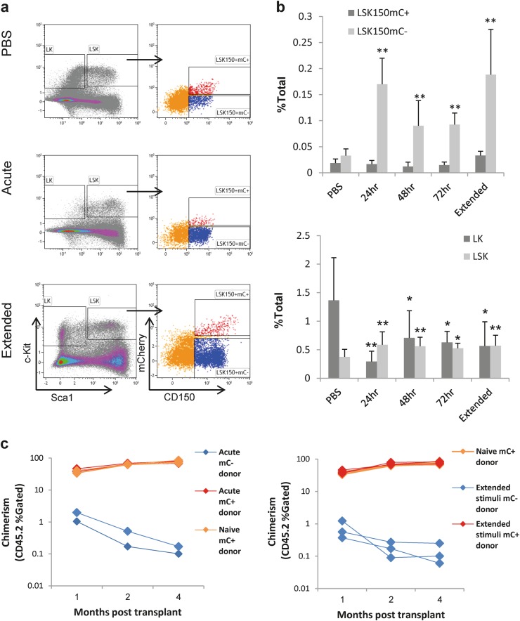
Acute and extended immune stimulation provokes conventional HSC markers. a Representative FACS plots, showing the staining of the Lineage−cKit+Sca1+ (LSK) compartment (left panels) and its dissection by CD150 and the Fgd5mCherry reporter (mC, right panels) under control conditions (top) and under acute (middle) and extended (bottom) pIC stimulation, 24 h post stimulation. b Quantification of the frequencies of indicated cell populations: Lineage-cKit+Sca1− (LK), Lineage-cKit+Sca1+ (LSK), LSKCD150+mC−, and LSKCD150+mC+. Histograms indicate mean frequency and standard deviation from the bone-marrow mononuclear cells (% of total). c LSKCD150+mC+ are functional HSCs. Chimerism (% of gated CD45.2 cells) over time following acute or extended pIC-stimulated donors. Data are from at least five mice per histogram; *p < 0.05, **p < 0.01
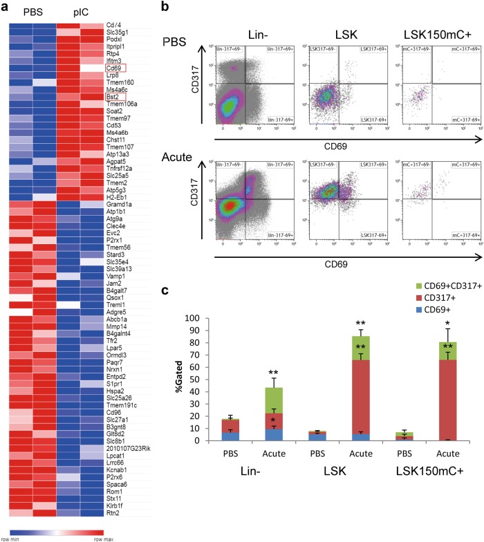
Surface markers of acutely activated HSCs. a A heatmap of 75 differentially expressed (DE) surface markers (GO:0016021, of 435 DE genes) under control (PBS) conditions and 24 h after an acute pIC stimulation. b Representative FACS plots showing the expression of CD69 (x-axis) and CD317 (Bts2, y-axis) in control conditions (PBS; top panels) and 24 h after an acute pIC stimulation (bottom panels). Plots are shown for the Lineage-negative (Lin−, left panels), Lineage−cKit+Sca1+ (LSK, middle panels), and LSKCD150+mCh+ (right panels) populations. c Quantification of the frequencies of indicated populations of CD69+CD317+ (green), CD317+CD69− (red), and CD69+CD317− (blue) cells. Histograms show averages and standard deviations of the control (PBS) and pIC-treated (Acute) cells; *p < 0.05, **p < 0.01
References
Publication types
MeSH terms
Substances
LinkOut - more resources
Full Text Sources
Other Literature Sources
Medical
Molecular Biology Databases
