Review
. 2018 Sep;15(9):543-554.
doi: 10.1038/s41569-018-0059-z.

Mitochondrial quality control mechanisms as molecular targets in cardiac ageing

Affiliations
Review

Mitochondrial quality control mechanisms as molecular targets in cardiac ageing

Anna Picca et al. Nat Rev Cardiol. 2018 Sep.

Abstract

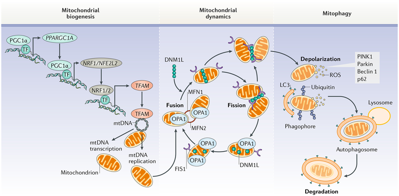
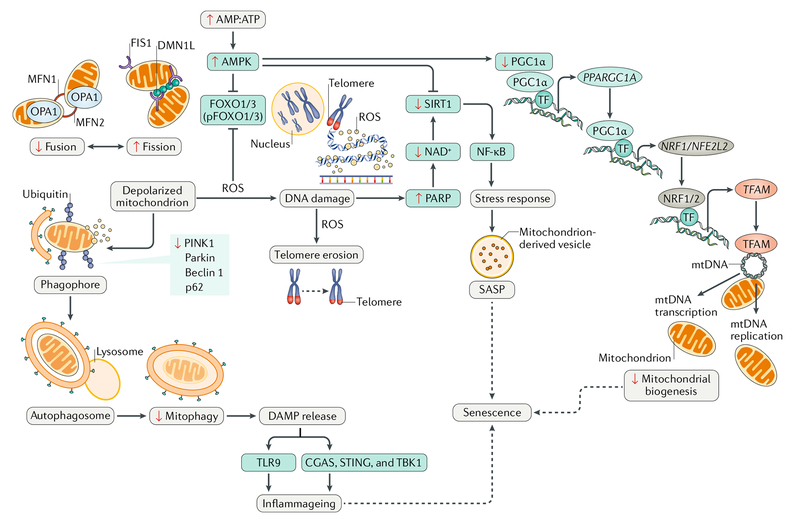
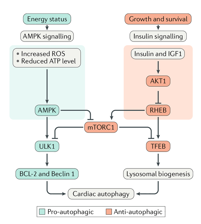
Cardiovascular disease is the leading cause of morbidity and mortality worldwide. Advancing age is a major risk factor for developing cardiovascular disease because of the lifelong exposure to cardiovascular risk factors and specific alterations affecting the heart and the vasculature during ageing. Indeed, the ageing heart is characterized by structural and functional changes that are caused by alterations in fundamental cardiomyocyte functions. In particular, the myocardium is heavily dependent on mitochondrial oxidative metabolism and is especially susceptible to mitochondrial dysfunction. Indeed, primary alterations in mitochondrial function, which are subsequently amplified by defective quality control mechanisms, are considered to be major contributing factors to cardiac senescence. In this Review, we discuss the mechanisms linking defective mitochondrial quality control mechanisms (that is, proteostasis, biogenesis, dynamics, and autophagy) to organelle dysfunction in the context of cardiac ageing. We also illustrate relevant molecular pathways that might be exploited for the prevention and treatment of age-related heart dysfunction.


Conflict of interest statement

Competing interests

The authors declare no competing interests.

Figures

Fig. 1 |
Fig. 1 |. Mitochondrial quality control pathways
Mitochondrial homeostasis is ensured through the coordination of mitochondrial biogenesis, dynamics, and autophagy. After appropriate stimuli (such as exercise stimulus), the upregulation of proliferator-activated receptor- γ co-activator 1α (PGC1α) and other transcription factors (TFs) activates the transcription of nuclear genes encoding mitochondrial proteins such as mitochondrial transcription factor A (TFAM). TFAM is then imported into mitochondria by the protein import machinery and reaches its final destination on mitochondrial DNA (mtDNA). Here, TFAM upregulates the expression of genes encoding electron transport chain subunits, resulting in increased oxygen consumption, ATP synthesis, and mitochondrial content. Changes in mitochondrial morphology are under the control of fusion (mitofusin 1 (MFN1), MFN2, and mitochondrial dynamin-like 120 kDa protein (OPA1)) and fission (dynamin 1-like protein (DNM1L) and mitochondrial fission 1 protein (FIS1)) proteins. These factors regulate mitochondrial turnover by facilitating the dilution and clearance of damaged organelles. Mitochondrial components are eventually recycled through a specialized autophagic pathway, known as mitophagy. LC3,microtubule-associated proteins 1A/1B light chain 3; NRF1, nuclear respiratory factor 1; NRF2, nuclear factor erythroid 2-related factor 2; p62, sequestosome 1; parkin, E3 ubiquitin-protein ligase parkin; PINK1, serine/threonine-protein kinase PINK1; ROS, reactive oxygen species.
Fig. 2 |
Fig. 2 |. Nucleus–mitochondrion crosstalk during cardiac ageing
The age-related surge in generation of reactive oxygen species (ROS) arising primarily from the accumulation of dysfunctional mitochondria causes nuclear DNA damage and activates 5′-AMP-activated protein kinase (AMPK) signalling. The latter, in turn, inhibits NAD-dependent protein deacetylase sirtuin 1 (SIRT1) activity. The decrease in NAD+ levels in the setting of oxidative stress further affects SIRT1, resulting in decreased levels of proliferator-activated receptor- γ co-activator 1α (PGC1α), leading to a decline in mitochondrial biogenesis; upregulation of nuclear factor- κB (NF- κB), leading to inflammation; and decreased expression and phosphorylation of forkhead box protein O1 (FOXO1) and FOXO3, transcription factors (TFs) that participate in cytoprotection. This multi-pathway derangement leads to cellular stress, induction of the senescence-associated secretory pathway (SASP), and senescence. CGAS, cGMP-AMP synthase; DAMP, damage-associated molecular pattern; DNM1L, dynamin 1-like protein; FIS1, mitochondrial fission 1 protein; MFN, mitofusin; mtDNA, mitochondrial DNA; NRF1, nuclear respiratory factor 1; NRF2, nuclear factor erythroid 2-related factor 2; OPA1, mitochondrial dynamin-like 120 kDa protein; p62, sequestosome 1; PARP, poly[ADP-ribose] polymerase; parkin, E3 ubiquitin-protein ligase parkin; PINK1, serine/threonine-protein kinase PINK1; STING, stimulator of interferon genes protein; TBK1, serine/threonine-protein kinase TBK1; TFAM, mitochondrial transcription factor A; TLR9, Toll-like receptor 9.
Fig. 3 |
Fig. 3 |. regulation of cardiac autophagy
Cardiomyocyte energy status regulates autophagy via metabolic signalling. Under substrate deficit conditions or oxidative stress, decreased ATP levels stimulate 5′-AMP-activated protein kinase (AMPK) activity and, therefore, autophagy via activation of serine/threonine-protein kinase ULK1 and downstream apoptosis regulator BCL-2 and beclin 1 through inhibition of autophagy suppressors (such as mechanistic target of rapamycin complex 1 (mTORC1)). At the same time, growth or cell survival stimuli activate insulin or insulin-like growth factor I (IGF1) signalling in cardiomyocytes, leading to the induction of the insulin–RACα serine/threonine-protein kinase (AKT1) pathway. Activation of GTP-binding protein RHEB results in inhibition of autophagy by autophagy suppressors (primarily mTORC1) and transcription factors related to lysosomal biogenesis (such as transcription factor EB (TFEB)). ROS, reactive oxygen species.

References

    1. Mozaffarian D et al. Heart disease and stroke statistics—2016 update. Circulation 133, e38–e360 (2016). - PubMed
    1. Chiao YA & Rabinovitch PS The aging heart. Cold Spring Harb. Perspect. Med. 5, a025148 (2015). - PMC - PubMed
    1. Zhang Y et al. Mitochondrial aldehyde dehydrogenase 2 accentuates aging-induced cardiac remodeling and contractile dysfunction: role of AMPK, Sirt1, and mitochondrial function. Free Radic. Biol. Med 71, 208–220 (2014). - PMC - PubMed
    1. Baris OR et al. Mosaic deficiency in mitochondrial oxidative metabolism promotes cardiac arrhythmia during aging. Cell Metab. 21, 667–677 (2015). - PubMed
    1. Lok NS & Lau CP Prevalence of palpitations, cardiac arrhythmias and their associated risk factors in ambulant elderly. Int. J. Cardiol 54, 231–236 (1996). - PubMed

Publication types