. 2018 Jul 24;8(1):11127.
doi: 10.1038/s41598-018-29444-0.

An acidic model pro-peptide affects the secondary structure, membrane interactions and antimicrobial activity of a crotalicidin fragment

Affiliations

An acidic model pro-peptide affects the secondary structure, membrane interactions and antimicrobial activity of a crotalicidin fragment

Nelson G O Júnior et al. Sci Rep. .

Abstract

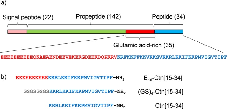
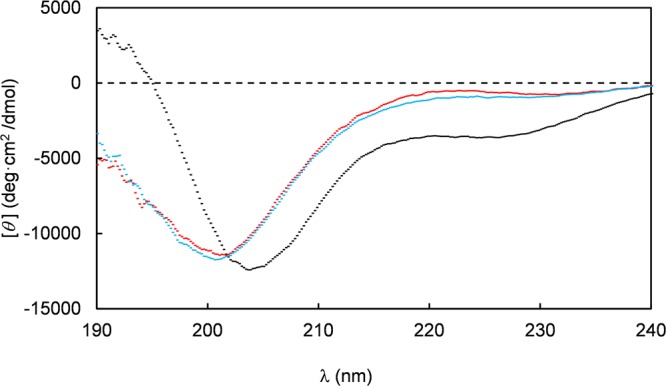
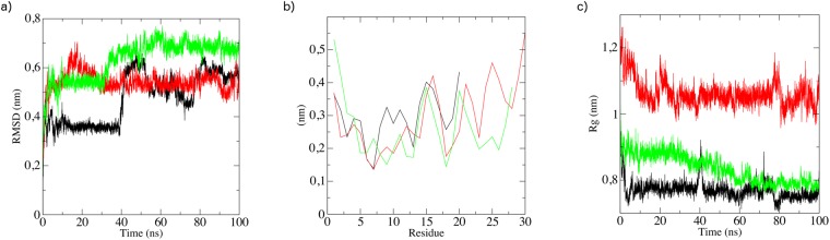
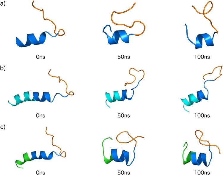
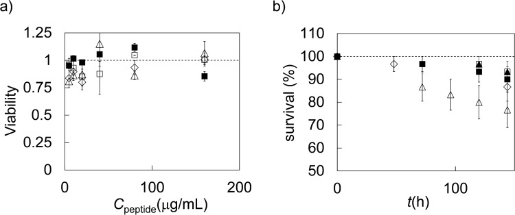
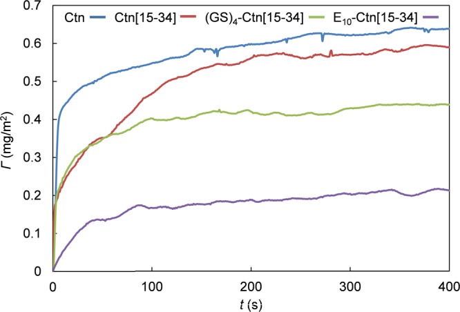
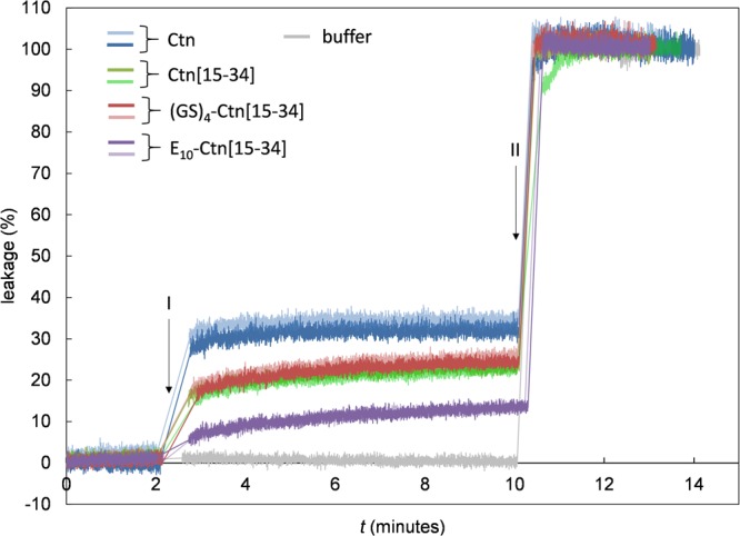
In order to study how acidic pro-peptides inhibit the antimicrobial activity of antimicrobial peptides, we introduce a simple model system, consisting of a 19 amino-acid long antimicrobial peptide, and an N-terminally attached, 10 amino-acid long acidic model pro-peptide. The antimicrobial peptide is a fragment of the crotalicidin peptide, a member of the cathelidin family, from rattlesnake venom. The model pro-peptide is a deca (glutamic acid). Attachment of the model pro-peptide only leads to a moderately large reduction in the binding to- and induced leakage of model liposomes, while the antimicrobial activity of the crotalicidin fragment is completely inhibited by attaching the model pro-peptide. Attaching the pro-peptide induces a conformational change to a more helical conformation, while there are no signs of intra- or intermolecular peptide complexation. We conclude that inhibition of antimicrobial activity by the model pro-peptide might be related to a conformational change induced by the pro-peptide domain, and that additional effects beyond induced changes in membrane activity must also be involved.


Conflict of interest statement

The authors declare no competing interests.

Figures

Figure 1
Figure 1
Crotalicidin-derived peptides used in this study. (a) Domain structure of crotalicidin pre-pro-peptide (Uniprot U5KJM4) in brackets: length of domains in numbers of amino acids. Flanking the peptide, the pro-peptide features a 35 amino acids long glutamic acid rich domain, whose sequence is also given in red (21 negatively charged residues, 6 positively charged residues), together with that of the full-length crotalicidin peptide in blue (15 positively charged residues). (b) Short model peptides inspired by full-length crotalicidin peptide and pro-peptide, used in this study. The Ctn[15–34] fragment has 7 positively charged residues.
Figure 2
Figure 2
Circular dichroism spectra of the peptides Ctn[15–34] (red), E10-Ctn[15–34] (black) and (GS)4-Ctn[15–34] (blue). Residue molar ellipticity [θ] in deg.cm2/dmol is plotted versus the wavelength (λ in nm). Measurements were performed at a peptide concentration of 0.2 μg.mL−1 in a 10 mM K2HPO4 50 mM Na2SO4 buffer, pH 7.4.
Figure 3
Figure 3
Molecular dynamics simulations in water (100 ns) for the peptides Ctn[15–34] (black curves), E10-Ctn[15–34] (red curves) and (GS)4-Ctn[15–34] (green curves), yielding the parameters (a) Root mean square deviation (RMSD) of single run, (b) root mean square fluctuations (RMSF) and (c) radius of gyration (Rg). Results are for single runs, three runs were done, for each peptide, with similar results.
Figure 4
Figure 4
Theoretical structures snapshots of (a) Ctn[15–34] (b) E10-Ctn[15–34] and (c) (GS)4-Ctn[15–34] during 100 ns of dynamics simulations (50 ns intervals). Domain colors: basic domain Ctn[15–34] – dark blue, hydrophobic domain Ctn[15–34] - orange, acidic E10 domain – light blue, control pro-peptide domain (GS)4 – green.
Figure 5
Figure 5
Toxicity of peptides Ctn[15–34] E10-Ctn[15–34] and GS4-Ctn[15–34]. (a) In vitro neutral red uptake assay for Caco-2 cells. Viability (fraction of viable cells) as compared to untreated Caco-2 cells as a function of peptide concentration. Diamonds: filled squares: PBS control, open squares: Ctn[15–34] open diamonds: E10-Ctn[15–34] open triangles: (GS)4-Ctn[15–34]. Error bars are standard deviations of replicate measurements. (b) In vivo toxicity of the peptides for Galleria mellonella larvae. Percentage surviving larvae as a function of time. Filled squares: H2O control, filled triangles: no treatment control, open triangles: Ctn[15–34] open diamonds: E10-Ctn[15–34] open squares: (GS)4-Ctn[15–34]. For each peptide a group of n = 15 larvae were used, error bars are standard deviations within the group.
Figure 6
Figure 6
Adsorption of the peptides Ctn, Ctn[15–34] E10-Ctn[15–34] and GS4-Ctn[15–34] to mixed DOPC:DOPG (7:3 molar ratio) supported lipid bilayers (SLB). Peptide mass Γ (mg/m2) adsorbing to the SLB as a function of time. Curves from top to bottom are for Ctn, Ctn[15–34] GS4-Ctn[15–34] and E10-Ctn[15–34] Peptide concentrations are 16 μg.mL−1 in 10 mM Tris 100 mM NaCl, pH 7.5.
Figure 7
Figure 7
Liposome leakage assay for the peptides Ctn, Ctn[15–34] E10-Ctn[15–34] and (GS)4-Ctn[15–34]. Percentage of leakage as a function of time t(s). Mixed DOPC:DOPG (7:3 molar ratio) liposomes with a diameter of 190 nm filled with calcein (70 mM inside the liposomes), at a lipid concentration of 50 μM, were exposed to 128 μg.mL−1 of the peptides. Peptides were injected at t = 2 min (I). At t = 10 min (II) a small amount of 10% (v/v) Triton X-100 was added to release all of the calcein. Two replicates are shown for each peptide with different colors as indicated in the figure.

References

    1. Neurath H, Walsh K. Role of proteolytic enzymes in biological regulation (A Review) A P. Natl. Acad. Sci. USA. 1976;73:3825–3832. doi: 10.1073/pnas.73.11.3825. - DOI - PMC - PubMed
    1. Salzman NH, et al. Enteric defensins are essential regulators of intestinal microbial ecology. Nat. Immunol. 2010;11:76–82. doi: 10.1038/ni.1825. - DOI - PMC - PubMed
    1. Shinnar AE, Butler KL, Park HJ. Cathelicidin family of antimicrobial peptides: proteolytic processing and protease resistance. Bioorg. Chem. 2003;31:425–436. doi: 10.1016/S0045-2068(03)00080-4. - DOI - PubMed
    1. van Ampting MTJ, et al. Intestinally secreted C-Type lectin Reg3b attenuates salmonellosis but not listeriosis in mice. Infect. Immun. 2012;80:1115–1120. doi: 10.1128/IAI.06165-11. - DOI - PMC - PubMed
    1. Cash HL, Whitham CV, Behrendt CL, Hooper LV. Symbiotic bacteria direct expression of an intestinal bactericidal lectin. Science. 2006;313:1126–1130. doi: 10.1126/science.1127119. - DOI - PMC - PubMed

Publication types

MeSH terms

LinkOut - more resources