. 2019 May;26(5):932-942.
doi: 10.1038/s41418-018-0171-y. Epub 2018 Jul 24.

STAT3 localizes to the ER, acting as a gatekeeper for ER-mitochondrion Ca2+ fluxes and apoptotic responses

Affiliations

STAT3 localizes to the ER, acting as a gatekeeper for ER-mitochondrion Ca2+ fluxes and apoptotic responses

Lidia Avalle et al. Cell Death Differ. 2019 May.

Abstract

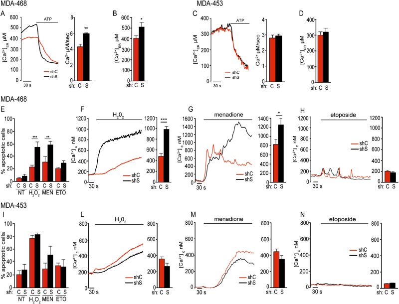
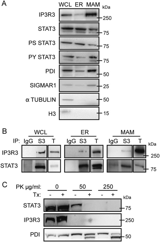
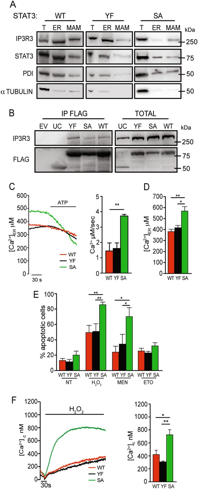
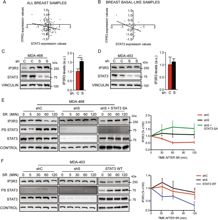
STAT3 is an oncogenic transcription factor exerting its functions both as a canonical transcriptional activator and as a non-canonical regulator of energy metabolism and mitochondrial functions. While both activities are required for cell transformation downstream of different oncogenic stimuli, they rely on different post-translational activating events, namely phosphorylation on either Y705 (nuclear activities) or S727 (mitochondrial functions). Here, we report the discovery of the unexpected STAT3 localization to the endoplasmic reticulum (ER), from where it modulates ER-mitochondria Ca2+ release by interacting with the Ca2+ channel IP3R3 and facilitating its degradation. The release of Ca2+ is of paramount importance for life/death cell decisions, as excessive Ca2+ causes mitochondrial Ca2+ overload, the opening of the mitochondrial permeability transition pore, and the initiation of the intrinsic apoptotic program. Indeed, STAT3 silencing enhances ER Ca2+ release and sensitivity to apoptosis following oxidative stress in STAT3-dependent mammary tumor cells, correlating with increased IP3R3 levels. Accordingly, basal-like mammary tumors, which frequently display constitutively active STAT3, show an inverse correlation between IP3R3 and STAT3 protein levels. These results suggest that STAT3-mediated IP3R3 downregulation in the ER crucially contributes to its anti-apoptotic functions via modulation of Ca2+ fluxes.


Conflict of interest statement

The authors declare that they have no conflict of interest.

Figures

Fig. 1
Fig. 1
Relevance of STAT3 in Ca2+ homeostasis and apoptotic response. MDA-MB-468 (a, b, eh) or MDA-MB-453 cells (c, d, in), silenced or not for STAT3 (shS, shSTAT3; shC, sh control) were used as indicated. a–d ER Ca2+ content and release. To induce Ca2+ release from ER, the cells were challenged with ATP, which evokes a rapid discharge from inositol 1,4,5-phosphate receptors (IP3Rs). a, c Representative traces are shown. ER calcium release (mean ± SEM) is quantified by the bars and expressed as μM/s. b, d The steady-state Ca2+ content. Bars are mean ± SEM of at least eight traces from three independent experiments. e, i Apoptosis upon treatment with hydrogen peroxide (H2O2), menadione (MEN), or etoposide (ETO), measured by cytofluorimetry of Annexin V/PI+ cells in the indicated cells. Bars represent the percentage of Annexin V/PI-positive cells (mean ± SEM from five independent experiments). fh, ln Cytoplasmic Ca2+ release was measured upon the indicated treatments. Bars are mean ± SEM of 12 measurements from three independent experiments. The asterisks indicate statistically significant differences. *P < 0.05; **P < 0.005; ***P < 0.001
Fig. 2
Fig. 2
STAT3 localizes to the ER and MAM compartments and interacts with IP3R3. a Whole-cell lysates from MDA-MB-468 cells were fractionated and analysed by Western blot. Representative of at least five independent experiments. b Co-immunoprecipitation of STAT3 and IP3R3. Whole-cell lysates or ER and MAM fractions were subjected to immunoprecipitation with anti-STAT3 antibodies (S3) or with control IgG, and blotted with the indicated antibodies. Representative of at least five independent experiments. WCL whole-cell lysate, ER endoplasmic reticulum, MAM mitochondrial-associated membranes, T total extract. c Whole-cell lysates from MDA-MB-468 cells were treated with the indicated amounts of Proteinase K (PK) in the presence or absence of 3% Triton-X 100 (Tx), and analysed by Western blot. Representative of five independent experiments
Fig. 3
Fig. 3
Characterization of STAT3-null MEFs replaced with WT or mutant STAT3. a Whole-cell lysates from STAT3 null MEF cells stably expressing wild type (WT), YF-, or SA-STAT3 were fractionated and analysed by Western blot. b HEK293 cells overexpressing IP3R3 were transiently transfected with flagged wild type or mutant STAT3 (WT, YF, SA) or with an unrelated control (UC), and immunoprecipitated with anti-flag antibodies followed by Western blot. EV empty vector. c, d ER Ca2+ release (c) and content (d) induced by treatment with ATP in MEFs expressing STAT3WT (red line), STAT3YF (black line) or STAT3SA (green line), measured as described in the legend to Fig. 1. Bars are mean ± SEM of 10 measurements from three independent experiments. e Apoptosis in response to hydrogen peroxide (H2O2), menadione (MEN), or etoposide (ETO), assessed in STAT3WT, SA, or YF MEFs by cytofluorimetric analysis of Annexin V/PI+ cells. Mean ± SEM from five independent experiments. NT, untreated. f Cytoplasmic Ca2+ release in STAT3WT, SA, or YF MEF cells upon H2O2 stimulation. Bars represent the mean ± SEM of 16 measurements. The asterisks indicate statistically significant differences. **P < 0.005; ***P < 0.001
Fig. 4
Fig. 4
STAT3-mediated regulation of IP3R3 protein levels in breast primary tumors and cell lines. a, b STAT3:IP3R3 anti-correlation in breast cancer patients from the CPTAC, TCGA Cancer Proteome Study of Breast Tissue dataset, considering all tumor subtypes (a, Pearson coefficient = −0.18, P = 0.06) or basal-like tumor samples (b, Pearson coefficient = −0.41, P = 0.04). c, d IP3R3 levels upon STAT3 silencing in MDA-MB-468 (c) or in MDA-MB-453 (d) cells. shS shSTAT3, shC sh control. Bars represent mean values ± SEM of four independent experiments. e, f IP3R3 levels upon starvation and serum restimulation (SR) in MDA-MB-468 (e) or MDA-MB-453 (f) cells silenced or not for STAT3 and overexpressing STAT3 SA or WT (shS, shSTAT3; shC, sh control). The graphs on the right show the relative quantification of IP3R3 levels from at least four independent experiments, as mean ± SEM. P values were calculated by two-way ANOVA. e, shC vs shS, P < 0.001; shC vs shS + STAT3 SA, P < 0.001; shS vs shS + STAT3 SA, ns. f, shC vs shS, ns; shC vs STAT3 WT, P < 0.05; shS vs STAT3 WT, P < 0.01

Comment in

References

    1. Yu H, Lee H, Herrmann A, Buettner R, Jove R. Revisiting STAT3 signalling in cancer: new and unexpected biological functions. Nat Rev Cancer. 2014;14:736–46. doi: 10.1038/nrc3818. - DOI - PubMed
    1. Yuan J, Zhang F, Niu R. Multiple regulation pathways and pivotal biological functions of STAT3 in cancer. Sci Rep. 2015;5:17663. doi: 10.1038/srep17663. - DOI - PMC - PubMed
    1. Pilati C, Amessou M, Bihl MP, Balabaud C, Nhieu JT, Paradis V, et al. Somatic mutations activating STAT3 in human inflammatory hepatocellular adenomas. J Exp Med. 2011;208:1359–66. doi: 10.1084/jem.20110283. - DOI - PMC - PubMed
    1. Waldmann TA. JAK/STAT pathway directed therapy of T-cell leukemia/lymphoma: inspired by functional and structural genomics. Mol Cell Endocrinol. 2017;451:66–70. doi: 10.1016/j.mce.2017.02.019. - DOI - PMC - PubMed
    1. Bromberg JF, Horvath CM, Besser D, Lathem WW, Darnell JE., Jr Stat3 activation is required for cellular transformation by v-src. Mol Cell Biol. 1998;18:2553–8. doi: 10.1128/MCB.18.5.2553. - DOI - PMC - PubMed

Publication types

MeSH terms