. 2017 Nov 7;42(4):209-214.
doi: 10.1080/01658107.2017.1392581. eCollection 2018 Aug.

Invasive Fungal Sinusitis Presenting as Acute Posterior Ischemic Optic Neuropathy

Affiliations

Invasive Fungal Sinusitis Presenting as Acute Posterior Ischemic Optic Neuropathy

Rafat Ghabrial et al. Neuroophthalmology. .

Abstract

Invasive fungal sinusitis causes painful orbital apex syndrome with ophthalmoplegia and visual loss; the mechanism is unclear. We report an immunocompromised patient with invasive fungal sinusitis in whom the visual loss was due to posterior ischaemic optic neuropathy, shown on diffusion-weighted MRI, presumably from fungal invasion of small meningeal-based arteries at the orbital apex. After intensive antifungal drugs, orbital exenteration and immune reconstitution, the patient survived, but we were uncertain if the exenteration helped. We suggest that evidence of acute posterior ischaemic optic neuropathy should be a contra-indication to the need for orbital exenteration in invasive fungal sinusitis.

Keywords: Invasive fungal sinusitis; orbital exenteration; posterior ischaemic optic neuropathy.


Figures

Figure 1a.
Figure 1a.
Contrast enhanced coronal T1 sequence (Day 13) demonstrates a communication between the right posterior ethmoid air cells and both the orbit and the anterior cranial fossa: fluid collection with enhancing margin extends from the sinuses to the inferior frontal lobe. The appearances are in keeping with an abscess. Note the herniation of the right medial rectus muscle through the surgical defect in the medial orbital wall. There is extensive pan-sinus opacification. There is no evidence of compression of the optic nerve at the orbital apex. ON=optic nerve; MR=medial rectus.
Figure 1b.
Figure 1b.
More anteriorly the thickened right medial rectus muscle extends medially into the right ethmoid air cells. SR=superior rectus; SO=superior oblique; MR=medial rectus; NS=nasal septum.
Figure 2a.
Figure 2a.
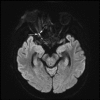
Diffusion weighted image (DWI) before orbital exenteration (Day 13) shows diffusion restriction (confirmed on ADC map) in the right optic nerve at the orbital apex and in the optic canal.
Figure 2b.
Figure 2b.
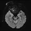
Diffusion weighted image (DWI) after orbital exenteration (Day 22) shows showing persistent diffusion restriction in the residual right optic nerve.
Figure 3a.
Figure 3a.
Ethmoid mucosa from FESS performed on 7 August shows angio-invasive fungal inflammation. Branching septate fungal hyphae are seen within infarcted vessels (arrow). PCR and culture were both positive in this specimen for Aspergillus fumigatus. DPAS special stain.
Figure 3b.
Figure 3b.
Deep orbital apex tissue. A few degenerate fungal hyphae were identified. The specimen was PCR positive but culture negative for Aspergillus. Preservation artefact is present as the specimen submitted first to microbiology for culture and PCR and to Anatomical Pathology only 48 hours later.

References

    1. Weinstein JM, Morris GL, ZuRhein GM, Gentry LR.. Posterior ischemic optic neuropathy due to Aspergillus fumigatus. J Clin Neuroophthalmol. 1989;9:7–13.bk_AQCmts2b - PubMed
    1. Overbeeke JJ, Sekhar LN.. Microanatomy of the blood supply to the optic nerve. Orbit. 2009;22:81–88. doi:10.1076/orbi.22.2.81.14316. - DOI - PubMed
    1. Sadda SR, Nee M, Miller NR, Biousse V, Newman NJ, Kouzis A. Clinical spectrum of posterior ischemic optic neuropathy. Am J Ophthalmol. 2001;132:743–750. doi:10.1016/S0002-9394(01)01199-0. - DOI - PubMed
    1. Däubler BF, Kamli A. Runge. Readout-segmented diffusion-weighted imaging in a critical anatomic area -diagnosing posterior ischemic optic neuropathy (PION). Rofo. 2014;186:1151–1152. doi:10.1055/s-0034-1366411. - DOI - PubMed
    1. Al-Zubidi N, Stevens S, Fung SH, Lee AG. Diffusion-weighted imaging in posterior ischemic optic neuropathy. Can J Ophthalmol. 2014;49:e21–5. doi:10.1016/j.jcjo.2013.11.003. - DOI - PubMed

LinkOut - more resources