Review
. 2015 Sep;1(1):23-29.
doi: 10.18383/j.tom.2015.00118.

Assessment of Pulmonary Arterial Hypertension by Magnetic Resonance Imaging

Affiliations
Review

Assessment of Pulmonary Arterial Hypertension by Magnetic Resonance Imaging

El-Sayed H Ibrahim et al. Tomography. 2015 Sep.

Abstract

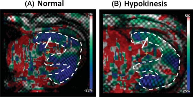
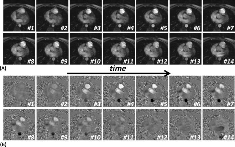
Pulmonary arterial hypertension (PAH) is characterized by elevated pulmonary artery pressure (PAP), altered pulmonary artery (PA) hemodynamics, and vessel wall characteristics that affect the right ventricular (RV) function. Magnetic resonance imaging (MRI) has recently been considered in PAH and has shown promising results for estimating PAP, measuring PA hemodynamic parameters, assessing PA vessel wall stiffness, and evaluating RV global and regional functions. In this article, we review various MRI techniques and image analysis methods for evaluating PAH, with an emphasis on the resulting images and how they are interpreted for both qualitatively and quantitatively assessing the PA and RV conditions.

Keywords: magnetic resonance imaging; pulmonary arterial hypertension; pulmonary artery; right ventricle.


Conflict of interest statement

Conflict of Interest: The authors declare no conflicts of interest.

Figures

Figure 1.
Figure 1.
Cine MR images. (A) Stack of parallel short-axis images covering both the right and left ventricles. (B) Sample images at different locations in the heart and times in the cardiac cycle.
Figure 2.
Figure 2.
Right ventricular (RV) hypertrophy in pulmonary arterial hypertension shown on cine MR images. Short-axis (top) and the corresponding 4-chamber (bottom) slices of a patient without hypertrophy (left) and patients with mild (middle) and severe (right) RV hypertrophy. Note the thick RV wall, which is translated into increased RV mass.
Figure 3.
Figure 3.
Right ventricular (RV) dilation. Four-chamber cine MR images showing a patient with nondilated RV (left) as well as patients with moderate (middle) and severe (right) RV dilation. Note the large chamber size, which leads to increased RV volume. The right atrium shows corresponding dilation in the provided examples.
Figure 4.
Figure 4.
Myocardial trabeculations in the right ventricle, as shown on short-axis cine MR images. Trabeculations are especially apparent at the midventricular (top) and apical (bottom) slices. Examples of normal (left), mild PAH (middle), and severe PAH (right) cases are shown.
Figure 5.
Figure 5.
Septal wall bending in pulmonary arterial hypertension (PAH). Basal short-axis cine MR images at early diastole showing normal septal wall shape (left) as well as mild, moderate, and severe leftward septal wall bowing at different PAH stages.
Figure 6.
Figure 6.
Right ventricular (RV) myocardial strain using myocardial tagging and harmonic phase analysis. Color-coded grid-tagged images showing longitudinal strain. The images show 2 examples of patients with normal RV contraction (A) and RV hypokinesis (B).
Figure 7.
Figure 7.
Strain encoding (SENC) MR images showing myocardial strain. Color-coded SENC images show through-plane longitudinal strain in patients with mild (A) and severe (B) pulmonary arterial hypertension. (C) Longitudinal right ventricular strain curves and early-diastolic strain rate at a remote RV site marked with ′x'.
Figure 8.
Figure 8.
Blood flow through the tricuspid valve during diastole, as shown on phase-contrast MR images acquired across the tricuspid valve with through-plane velocity encoding. (A) Reference magnitude image and a sequence of phase images at consecutive timepoints in the cardiac cycle. The tricuspid valve (arrow) opens, and the blood flows from the right atrium to the right ventricle. (B, C) Tricuspid blood flow curves from 2 patients with an early-to-atrial filling ratio greater than 1 (B) and less than 1 (C), reflecting normal and abnormal diastolic functions, respectively.
Figure 9.
Figure 9.
Pulmonary artery (PA) dilation, as shown on oblique sagittal anatomical images. Oblique images show a patient with normal PA size (left), as well as patients with moderate (middle) and severe (right) PA dilation. MPA, main PA.
Figure 10.
Figure 10.
Consecutive frames of magnitude (top) and phase (bottom) velocity-encoded images acquired at the pulmonary valve level during systole. The images are used to draw the flow curve, from which pulmonary artery hemodynamic parameters are measured.
Figure 11.
Figure 11.
Pulmonary flow pattern and hemodynamic parameters based on velocity-encoding MRI. The flow curve and parameters are calculated from the phase-encoded images acquired perpendicular to the flow direction at the pulmonary valve level.
Figure 12.
Figure 12.
Pulmonary artery (PA) distensibility and pulse wave velocity (PWV), as measured using the flow-area method applied on velocity-encoded MRIs. (A) Successive frames of magnitude (top) and the corresponding phase (bottom) images showing early-systolic blood flow in the PA (arrow) in a patient with mild pulmonary arterial hypertension (PAH). Noticeable increases in the PA cross-sectional area and blood flow are observed. (B) PWV is calculated from the images in (A) as the ratio of change in blood flow to change in PA area. Images shown in (C) and (D) are similar to (A) and (B) in a patient with severe PAH (slight change in PA cross-sectional area, which results in a large PWV).

References

    1. Alassas K, Mergo P, Ibrahim el S, Burger C, Safford R, Parikh P, Shapiro B. Cardiac MRI as a diagnostic tool in pulmonary hypertension. Future Cardiol. 2014;10(1):117–130.. - PubMed
    1. Wang N, Hu X, Liu C, Ali B, Guo X, Liu M, Peng X, Yang Y. A systematic review of the diagnostic accuracy of cardiovascular magnetic resonance for pulmonary hypertension. Can J Cardiol. 2014;30(4):455–463. - PubMed
    1. Swift AJ, Wild JM, Nagle SK, Roldán-Alzate A, François CJ, Fain S, Johnson K, Capener D, van Beek EJ, Kiely DG, Wang K, Schiebler ML. Quantitative magnetic resonance imaging of pulmonary hypertension: a practical approach to the current state of the art. J Thorac Imaging. 2014;29(2):68–79. - PMC - PubMed
    1. Benza R, Biederman R, Murali S, et al. . Role of cardiac magnetic resonance imaging in the management of patients with pulmonary arterial hypertension. J Am Coll Cardiol. 2008;52:1683–1692. - PubMed
    1. Kovacs G, Reiter G, Reiter U, Rienmüller R, Peacock A, Olschewski H. The emerging role of magnetic resonance imaging in the diagnosis and management of pulmonary hypertension. Respiration. 2008;76:458–470. - PubMed

LinkOut - more resources