Review
. 2018 Aug;12(4):361-373.
doi: 10.1007/s11684-018-0656-6. Epub 2018 Jul 24.

Epithelial-to-mesenchymal transition in cancer: complexity and opportunities

Affiliations
Review

Epithelial-to-mesenchymal transition in cancer: complexity and opportunities

Yun Zhang et al. Front Med. 2018 Aug.

Abstract

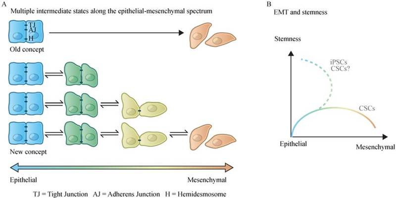
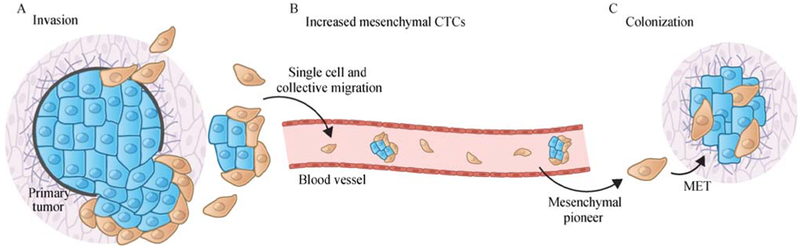
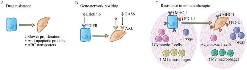
The cell-biological program termed the epithelial-to-mesenchymal transition (EMT) plays an important role in both development and cancer progression. Depending on the contextual signals and intracellular gene circuits of a particular cell, this program can drive fully epithelial cells to enter into a series of phenotypic states arrayed along the epithelial-mesenchymal phenotypic axis. These cell states display distinctive cellular characteristics, including stemness, invasiveness, drug-resistance and the ability to form metastases at distant organs, and thereby contribute to cancer metastasis and relapse. Currently we still lack a coherent overview of the molecular and biochemical mechanisms inducing cells to enter various states along the epithelial-mesenchymal phenotypic spectrum. An improved understanding of the dynamic and plastic nature of the EMT program has the potential to yield novel therapies targeting this cellular program that may aid in the management of high-grade malignancies.

Keywords: cancer; cancer stem cell; epithelial-to-mesenchymal transition; metastasis.


Figures

Fig. 1
Fig. 1
The dynamic and plastic nature of the EMT program. (A) Rather than a unidirectional binary switch between two distinct cell states, accumulating evidence suggests that the epithelial-to-mesenchymal transition (EMT) program generates a spectrum of different intermediate cell states between the extreme epithelial and mesenchymal endpoints. (B) Activation of EMT program is associated with the entrance into stem cell programs, though in certain contexts, constitutive activation of an EMT program in carcinoma cells leads to the loss of stem-like properties. Cancer cells undergone a sequential EMT-MET reprogramming could be very different from the original epithelial cells in the primary tumor. When reprogramming somatic cells into induced pluripotent stem cells (iPSCs), sequential introduction of Yamanaka factors in a specific order (first OCT-4 with KLF4, then c-MYC, and finally SOX2), rather than the simultaneous exposure, has been found to significantly improve the reprogramming efficiency. In this specific protocol, a sequential EMT-MET state change has been observed, showing an intermediate state with upregulated EMT-TFs and enhanced mesenchymal characteristics before entering the epithelial pluripotent state [112]. It is plausible that a similar sequential EMT-MET transition could generate cancer cells with increased stemness and the ability to form macro-metastatic colonies.
Fig. 2
Fig. 2
The EMT program facilitates multiple steps of the invasion-metastatic cascade. (A) At the primary tumor site, induction of an EMT program allows carcinoma cells to lose cell-cell junctions, degrade local basement membrane via elevated expression of various matrix-degrading enzymes and supports cancer cell dissemination in both the “single cell” and “collective migration” modes. (B) Many circulating tumor cells (CTCs), representing carcinoma cells that have entered into the vasculature and may thereafter be capable of seeding new metastatic colonies at distant anatomical sites, display partial EMT activation with co-expression both epithelial and mesenchymal markers. Moreover, mesenchymal CTCs have been found to be significantly enriched in cancer patients with refractory or progressive disease [45]. (C) At the colonization step, robust outgrowth of macro-metastases, at least in some destination organ sites, requires the reversion of EMT program and the associated gain of epithelial characteristics.
Fig. 3
Fig. 3
EMT confers therapeutic resistance. (A) EMT confers multidrug resistance on cancer cells. EMT-induced multidrug resistance involves a number of mechanisms, including a slow proliferation rate, elevated expression of anti-apoptotic proteins, and upregulation of ATP binding cassette (ABC) transporters that mediate drug efflux. (B) The E-to-M transition may induce cancer cells into novel phenotypic states and make certain therapeutic targets dispensable for continued cell viability. For example, the E-to-M transition switches the dependence of carcinoma cells from the EGFR to the AXL receptor tyrosine kinase in non-small-cell lung cancer cells, thereby yielding resistance to EGFR-targeted therapy. (C) The EMT program contributes to the establishment of an immunosuppressive tumor microenvironment and confers resistance to immunotherapies. In a cell-autonomous manner, induction of EMT program in carcinoma cells downregulates MHC-I molecules and β2-microglobulin while upregulating PD-L1. In addition, induction of EMT program leads to various non-cell-autonomous changes, remodeling the tumor microenvironment by recruiting M2 (pro-tumorigenic) macrophages and T-regs, and suppressing the infiltration of cytotoxic T cells.

References

    1. Nieto MA, Huang RYJ, Jackson RA, Thiery JP. EMT: 2016. Cell 2016; 166(1): 21–45 - PubMed
    1. Polyak K, Weinberg RA. Transitions between epithelial and mesenchymal states: acquisition of malignant and stem cell traits. Nat Rev Cancer 2009; 9(4): 265–273 - PubMed
    1. Greenburg G, Hay ED. Epithelia suspended in collagen gels can lose polarity and express characteristics of migrating mesenchymal cells. J Cell Biol 1982; 95(1): 333–339 - PMC - PubMed
    1. Lim J, Thiery JP. Epithelial-mesenchymal transitions: insights from development. Development 2012; 139(19): 3471–3486 - PubMed
    1. Carver EA, Jiang R, Lan Y, Oram KF, Gridley T. The mouse snail gene encodes a key regulator of the epithelial-mesenchymal transition. Mol Cell Biol 2001; 21(23): 8184–8188 - PMC - PubMed