. 2018 Jul 25;10(8):243.
doi: 10.3390/cancers10080243.

Activation of ERBB4 in Glioblastoma Can Contribute to Increased Tumorigenicity and Influence Therapeutic Response

Affiliations

Activation of ERBB4 in Glioblastoma Can Contribute to Increased Tumorigenicity and Influence Therapeutic Response

Jacqueline F Donoghue et al. Cancers (Basel). .

Abstract

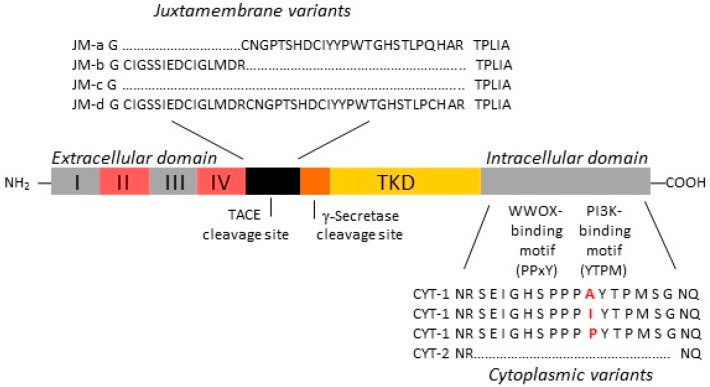
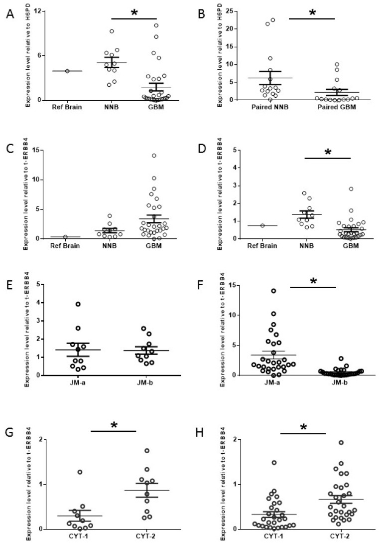
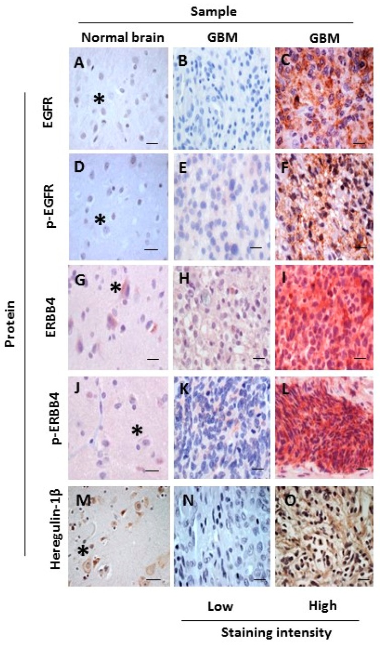
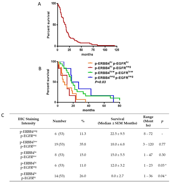
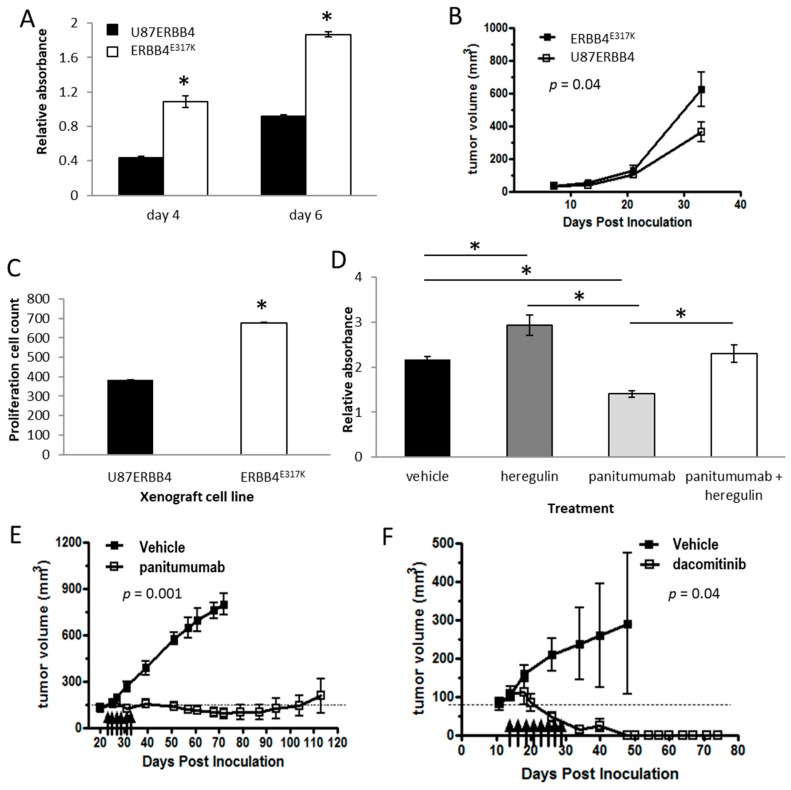
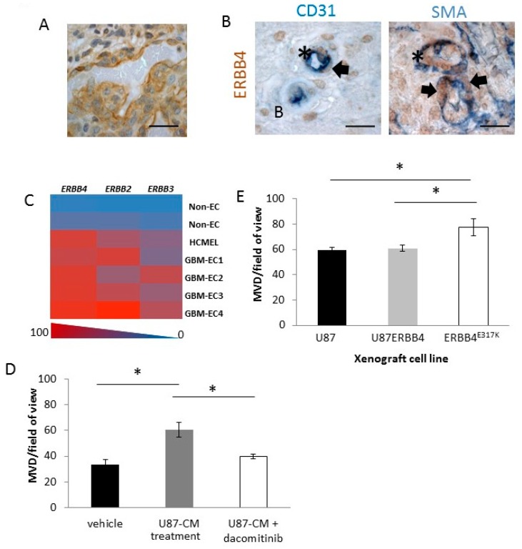
Glioblastoma (GBM) is often resistant to conventional and targeted therapeutics. ErbB2 Receptor Tyrosine Kinase 4 (ERBB4) is expressed throughout normal brain and is an oncogene in several pediatric brain cancers; therefore, we investigated ERBB4 as a prognostic marker and therapeutic target in GBM. Using RT-qPCR, we quantified mRNA encoding total ERBB4 and known ERBB4 variants in GBM and non-neoplastic normal brain (NNB) samples. Using immunohistochemistry, we characterized the localization of total and phosphorylated ERBB4 (p-ERBB4) and EGFR protein in archived GBM samples and assessed their association with patient survival. Furthermore, we evaluated the effect of ERBB4 phosphorylation on angiogenesis and tumorigenicity in GBM xenograft models. Total ERBB4 mRNA was significantly lower in GBM than NNB samples, with the juxtamembrane JM-a and cytoplasmic CYT-2 variants predominating. ERBB4 protein was ubiquitously expressed in GBM but was not associated with patient survival. However, high p-ERBB4 in 11% of archived GBM samples, independent of p-EGFR, was associated with shorter patient survival (12.0 ± 3.2 months) than was no p-ERBB4 (22.5 ± 9.5 months). Increased ERBB4 activation was also associated with increased proliferation, angiogenesis, tumorigenicity and reduced sensitivity to anti-EGFR treatment in xenograft models. Despite low ERBB4 mRNA in GBM, the functional effects of increased ERBB4 activation identify ERBB4 as a potential prognostic and therapeutic target.

Keywords: EGFR; ERBB4; GBM; prognosis; therapy.


Conflict of interest statement

Panitumumab was supplied to TGJ by Amgen, and dacomitinib was supplied to TGJ and NGG by Pfizer through a grant-in-aid.

Figures

Figure 1
Figure 1
Schematic representation of ERBB4 juxtamembrane (JM-a,b,c,d) and cytoplasmic (CYT-1,2) variant sequences. JM-a- and JM-d-containing variants are cleaved by tumour necrosis factor converting enzyme (TACE), releasing the extracellular domain. Further cleavage occurs at the γ-secretase site of the JM-a- and JM-d- containing variants, releasing a soluble intracellular fragment containing the tyrosine kinase domain (TKD) and the cytoplasmic tail from the inner cell membrane. JM-b and JM-c have no TACE cleavage site; therefore, variants containing either of these remain as a full-length receptor. CYT-1-containing variants include a 16 amino acid sequence that is absent from CYT-2-containing variants and contains WW domain-containing oxidoreductase (WWOX)- and PI3K-binding motifs. Amino acid sequences were generated from NCBI Nucleotide database.
Figure 2
Figure 2
Total ERBB4 and ERBB4 variant expression. ERBB4 expression in 10 non-neoplastic normal brain (NNB), 28 glioblastoma (GBM), and 15 paired NNB and GBM samples. Expression levels were determined by RT-qPCR; total ERBB4 levels were standardized to H6PD mRNA, and variant levels were standardized to total ERBB4 mRNA levels. All measurements were made simultaneously but are displayed separately to aid comparison. Reference brain (Ref Brain) was a pooled brain sample from 7 donors (Ambion, Thermo-Fisher, Waltham, MA, USA) used for comparison. Total ERBB4 mRNA expression in Ref Brain, NNB and GBM samples (A). Total ERBB4 mRNA expression in paired NNB and GBM samples (B). Expression of JM-a (C) and JM-b (D) variants of ERBB4 in Ref Brain, NNB and GBM samples. Comparison of JM-a and JM-b expression levels in NNB (E) and GBM (F). (Note that the data shown in (E,F) are rescaled from (C) and (D).) CYT-1 and CYT-2 expression levels in NNB (G) and GBM (H). Experiments were performed in triplicate (* p < 0.05, Student’s t-test).
Figure 3
Figure 3
Representative IHC images of occipital regions in an NNB sample and a GBM sample. Samples were stained for EGFR (AC), p-EGFR (DF), ERBB4 (GI), p-ERBB4 (JL) and heregulin-1β protein (MO). Images are representative of high and low staining intensity of ERBB proteins (stained with VECTOR® Red Alkaline Phosphatase (red), Burlingame, CA, USA) and of heregulin-1β protein (stained with 3,3′-Diaminobenzidine (DAB) (brown); nuclei were counterstained with hematoxylin (blue). Asterisks denote neurons. Scale bars, 25 µm.
Figure 4
Figure 4
Comparative survival of patients with GBM. The overall survival for the 53 patients with GBM for whom survival data were available ranged from 1 to 120 months (A). Graphical (B) and tabulated (C) survival outcomes associated with patient samples with high (hi), low (low) and no (neg) p-ERBB4 or p-EGFR staining by IHC. Combinations not shown in the table were not identified. (* p < 0.05 vs. p-ERBB4neg p-EGFRneg samples, Student’s t-test).
Figure 5
Figure 5
Activated ERBB4 and cell growth potential. The proliferation of U87MG cells infected with retrovirus carrying wild-type ERBB4 (U87ERBB4) was compared with hyper-activated ERBB4-expressing cells (ERBB4E317K) in vitro using an MTS proliferation assay at day 4 and 6 following drug addition (A), in vivo using xenograft tumor growth measured over time (B) and ex vivo using IHC analysis of Ki67 staining (C). MTS proliferation assay of SF767 cells treated with heregulin-1β (40 ng/mL), panitumumab (10 µg/mL) or both (D). Tumor volumes of SF767 xenografts treated with panitumumab (E) or dacomitinib (F). Arrows denote treatment times. Bars indicate mean values ± SEM (n = 4). Asterisks indicate bars that are significantly different from each other (* p < 0.05, Student’s t-test). All in vitro experiments were performed in triplicate. The in vivo tumor data include six repeats per group.
Figure 6
Figure 6
Angiogenic role of ERBB4. (A,B) p-ERBB4 expression on the vasculature of the GBM glomeruloid structure identified with DAB (brown), with nuclei counterstained with hematoxylin (A). IHC double stains demonstrated p-ERBB4 (brown) expression on endothelial cells (CD31, blue) and on pericytes (SMA, blue) in GBM patient samples (B). All IHC was performed on four biological replicates. Arrows point to pericytes, and asterisks identify endothelial cells. Scale bars, 25 µm. (C) Heat map of ERBB4, ERBB3 and ERBB2 mRNA expression of endothelial cells isolated from four samples of GBM (GBM-EC), one normal brain endothelial cell line (HCMEC) and two non-endothelial cell lines (non-EC), as measured by RT-qPCR. RT-qPCR was performed on triplicate samples. (D,E) MVD counts from subcutaneous agar plugs containing U87MG conditioned medium (U87-CM) following treatment with dacomitinib (D); representative images are shown in Figure S9A. MVD counts for U87MG, U87ERBB4 and ERBB4E317K xenografts (E); representative images are shown in Figure S9B. Bars indicate mean values ± SEM (n = 4). Asterisks indicate bars that are significantly different from each other (p < 0.05, one-way ANOVA).

References

    1. Maher E.A., Furnari F.B., Bachoo R.M., Rowitch D.H., Louis D.N., Cavenee W.K., DePinho R.A. Malignant glioma: Genetics and biology of a grave matter. Genes Dev. 2001;15:1311–1333. doi: 10.1101/gad.891601. - DOI - PubMed
    1. Wen P.Y., Kesari S. Malignant gliomas in adults. N. Engl. J. Med. 2008;359:492–507. doi: 10.1056/NEJMra0708126. - DOI - PubMed
    1. Inda M.M., Bonavia R., Mukasa A., Narita Y., Sah D.W., Vandenberg S., Brennan C., Johns T.G., Bachoo R., Hadwiger P., et al. Tumor heterogeneity is an active process maintained by a mutant EGFR-induced cytokine circuit in glioblastoma. Genes Dev. 2010;24:1731–1745. doi: 10.1101/gad.1890510. - DOI - PMC - PubMed
    1. Van den Bent M.J., Brandes A.A., Rampling R., Kouwenhoven M.C., Kros J.M., Carpentier A.F., Clement P.M., Frenay M., Campone M., Baurain J.F., et al. Randomized phase II trial of erlotinib versus temozolomide or carmustine in recurrent glioblastoma: EORTC brain tumor group study 26034. J. Clin. Oncol. 2009;27:1268–1274. doi: 10.1200/JCO.2008.17.5984. - DOI - PMC - PubMed
    1. Brown P.D., Krishnan S., Sarkaria J.N., Wu W., Jaeckle K.A., Uhm J.H., Geoffroy F.J., Arusell R., Kitange G., Jenkins R.B., et al. Phase I/II trial of erlotinib and temozolomide with radiation therapy in the treatment of newly diagnosed glioblastoma multiforme: North central cancer treatment group study N0177. J. Clin. Oncol. 2008;26:5603–5609. doi: 10.1200/JCO.2008.18.0612. - DOI - PMC - PubMed