Review
. 2018 Oct 6:34:545-568.
doi: 10.1146/annurev-cellbio-100617-062636. Epub 2018 Jul 25.

Intercellular Spread of Protein Aggregates in Neurodegenerative Disease

Affiliations
Review

Intercellular Spread of Protein Aggregates in Neurodegenerative Disease

Albert A Davis et al. Annu Rev Cell Dev Biol. .

Abstract

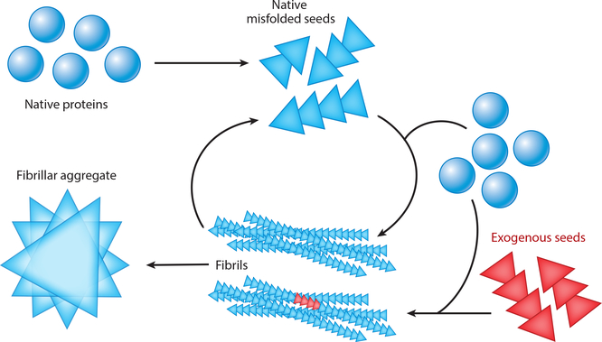
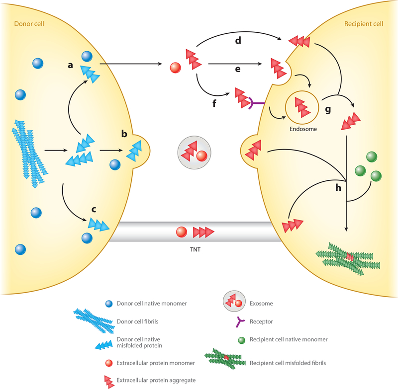
Most neurodegenerative diseases are characterized by the accumulation of protein aggregates, some of which are toxic to cells. Mounting evidence demonstrates that in several diseases, protein aggregates can pass from neuron to neuron along connected networks, although the role of this spreading phenomenon in disease pathogenesis is not completely understood. Here we briefly review the molecular and histopathological features of protein aggregation in neurodegenerative disease, we summarize the evidence for release of proteins from donor cells into the extracellular space, and we highlight some other mechanisms by which protein aggregates might be transmitted to recipient cells. We also discuss the evidence that supports a role for spreading of protein aggregates in neurodegenerative disease pathogenesis and some limitations of this model. Finally, we consider potential therapeutic strategies to target spreading of protein aggregates in the treatment of neurodegenerative diseases.

Keywords: neurodegenerative disease; protein aggregate; seeding; spreading.


Figures

Figure 1
Figure 1
Schematic representation of templated assembly of protein aggregates from spontaneous assembly of misfolded native protein or introduction of exogenous seeds. Briefly, native proteins (blue circles) may spontaneously misfold and subsequently propagate their misfolded conformation by templated recruitment of additional native proteins. These misfolded proteins can form oligomeric seeds (blue triangles). Misfolded seeds can continue to recruit native protein and self-assemble to form long, β-sheet-rich fibrils that ultimately form fibrillar aggregates. Alternatively, exogenous seeds (red triangles), either generated by another cell in a disease state or artificially introduced in experiments, may enter a cell and similarly recruit native proteins to form aggregates. Fibrils may also fragment into oligomers that may spur additional fibril formation within the same cell or that may spread to other cells to propagate protein misfolding and aggregation.
Figure 2
Figure 2
Potential mechanisms for exit of native and misfolded proteins from a donor cell and entry of exogenous seeds into a recipient cell. Proteins from a donor cell (blue) can exit by one of at least three pathways: (a) direct secretion of the free protein into the extracellular space by a currently unknown mechanism, (b) secretion in exosomes, or (c) direct transfer through tunneling nanotubes (TNTs). While these mechanisms have been reported to occur under physiological conditions, the role of secretion/release of normally intracellular proteins in brain function is unclear. Once in the extracellular space, free exogenous seeds (red) may then be taken up by a recipient cell by (d) direct insertion through the plasma membrane, (e) fluid-phase endocytosis or macropinocytosis, or (f) receptor-mediated endocytosis. If taken up through an endosome (g), exogenous seeds can escape the vesicle into the cytosol through a mechanism that is unclear. Seeds delivered to a recipient cell by exosomes or TNTs are directly released into the intracellular space. Once exogenous seeds are in the cytosol of the recipient cell (h), they may begin recruiting native monomer (green) and initiate templated misfolding, leading to the formation of oligomers/fibrils and eventually aggregates in the recipient cell.
Figure 3
Figure 3
Transfer of neurodegenerative disease–associated proteins between central-nervous-system cell types. The majority of proteins that aggregate in neurodegenerative diseases originate in neurons, and aggregates can be transferred to multiple cell types, including other neurons, astrocytes, microglia, and oligodendrocytes. Examples of transfer of specific proteins between particular cell types are indicated. Abbreviations: αSyn, alpha synuclein; Aβ, amyloid-β peptide; Htt, huntingtin; PrPSc, misfolded prion protein; SOD1, superoxide dismutase 1; TDP-43, 43-kDa TAR DNA-binding protein.

References

    1. Abounit S, Bousset L, Loria F, Zhu S, de Chaumont F, et al. 2016a. Tunneling nanotubes spread fibrillar α-synuclein by intercellular trafficking of lysosomes. EMBO J. 35:2120–38 - PMC - PubMed
    1. Abounit S, Wu JW, Duff K, Victoria GS, Zurzolo C. 2016b. Tunneling nanotubes: a possible highway in the spreading of tau and other prion-like proteins in neurodegenerative diseases. Prion 10:344–51 - PMC - PubMed
    1. Affiris AG. 2014. Studies assessing tolerability and safety of AFFITOPE® PD03A in patients with early Parkinson’s disease (AFF011). Rep. NCT02267434, US Natl. Libr. Med., Natl. Inst. Health, Washington, DC: https://clinicaltrials.gov/ct2/show/NCT02267434
    1. Alzheimer A 1907. Über eine eigenartige Erkrankung der Hirnrinde. Allg. Z. Psychiatr 64:146–48
    1. Andrade-Moraes CH, Oliveira-Pinto AV, Castro-Fonseca E, da Silva CG, Guimaraes DM, et al. 2013. Cell number changes in Alzheimer’s disease relate to dementia, not to plaques and tangles. Brain 136:3738–52 - PMC - PubMed

Publication types

MeSH terms

Substances