Review
. 2019 Jan 6:59:129-148.
doi: 10.1146/annurev-pharmtox-010617-052509. Epub 2018 Jul 25.

Challenges in Orphan Drug Development: Identification of Effective Therapy for Thyroid-Associated Ophthalmopathy

Affiliations
Review

Challenges in Orphan Drug Development: Identification of Effective Therapy for Thyroid-Associated Ophthalmopathy

Terry J Smith. Annu Rev Pharmacol Toxicol. .

Abstract

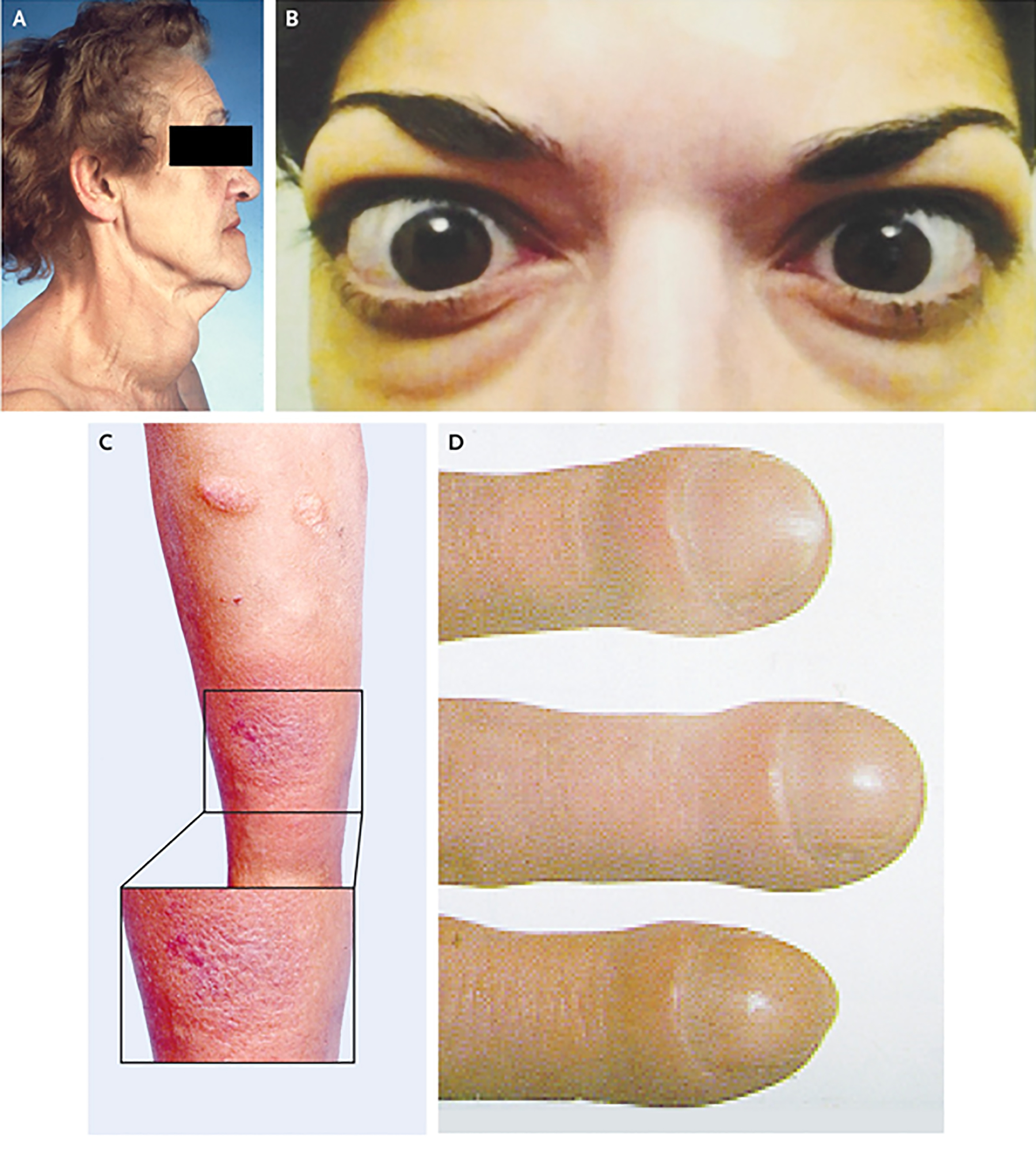
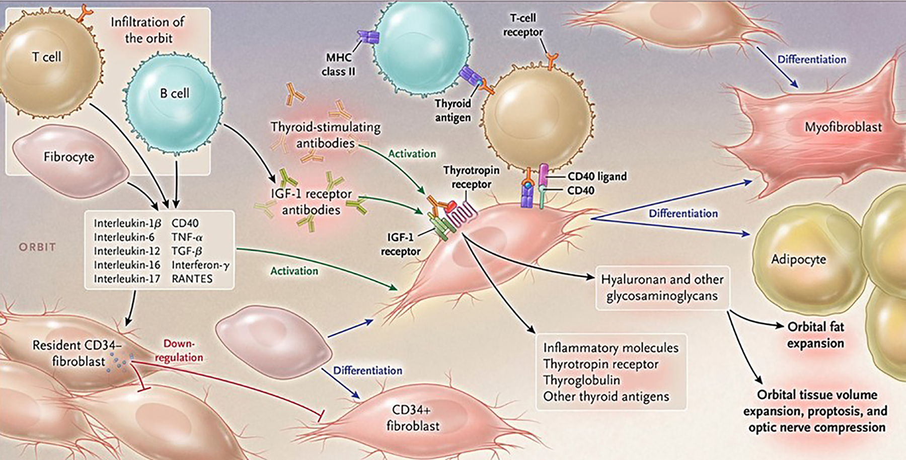
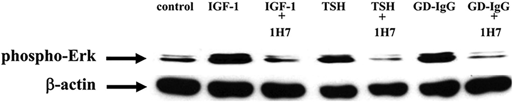
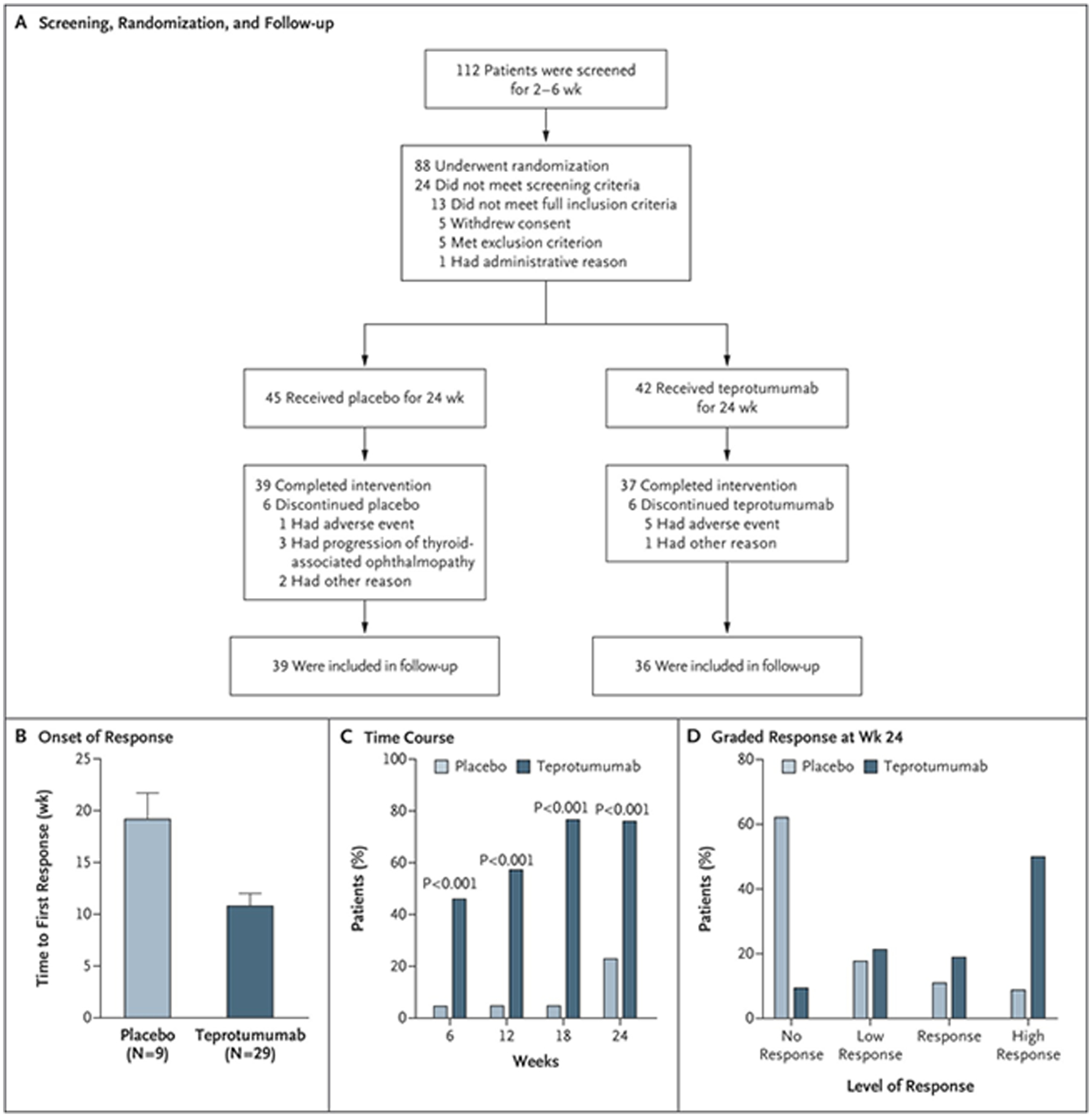
Thyroid-associated ophthalmopathy (TAO), the ocular manifestation of Graves' disease, is a process in which orbital connective tissues and extraocular muscles undergo inflammation and remodeling. The condition seems to result from autoimmune responses to antigens shared by the thyroid and orbit. The thyrotropin receptor (TSHR), expressed at low levels in orbital tissues, is a leading candidate antigen. Recent evidence suggests that another protein, the insulin-like growth factor-I receptor (IGF-IR), is overexpressed in TAO, and antibodies against IGF-IR have been detected in patients with the disease. Furthermore, TSHR and IGF-IR form a physical and functional complex, and signaling initiated at TSHR requires IGF-IR activity. Identification of therapy for this rare disease has proven challenging and currently relies on nonspecific and inadequate agents, thus representing an important unmet need. A recently completed therapeutic trial suggests that inhibiting IGF-IR activity with a monoclonal antibody may be an effective and safe treatment for active TAO.

Keywords: Graves’ disease; autoimmune; monoclonal antibody; signaling.


Conflict of interest statement

Conflicts

The author has been issued patents related to the detection of antibody-mediated inflammatory auto-immune disorders (US 6936426), the diagnosis and therapy of antibody-mediated inflammatory autoimmune disorders (US 7998681 and US 8153121), and diagnostic methods related to Graves’ disease and other autoimmune disorders (US 8178304). He was previously a paid consultant for River Vision.

Figures

Figure 1.
Figure 1.
Clinical appearance of Graves’ disease. Panel A demonstrates goiter (thyroid enlargement) frequently developing in the disease. Panel B shows moderate to severe thyroid-associated ophthalmopathy, including bilateral proptosis, lid retraction and periorbital edema. Panel C contains the image of plaque pretibial myxedema. Panel D demosntrates thyroid acropachy. From N. Engl. J. Med, Smith T.J. and Hegedus L., Graves’ Disease, 375; 1552–1565. Copyright © (2016) Massachusetts Medical Society. Reprinted with permission.
Figure 2.
Figure 2.
Pathogenesis of thyroid-associated ophthalmopathy. Orbital tissues infiltrated by B and T cells and CD34+ fibrocytes become activated in the disease process by virtue of cell-cell cross talk. Fibrocytes trafficked from bone marrow promiscuously express “thyroid-specific” proteins, including the thyrotropin receptor, thyroglobulin, and other inflammatory genes. They undergo differentiation into myofibroblasts or adipocytes, a consequence of their molecular environment. CD34+ fibroblasts, derived from the fibrocytes take up residence with “garden variety” CD34 fibroblasts. In aggregate, these fibroblasts generate interleukins 1β, 6, 8, 10, 16, IL-1 receptor antagonists, tumor necrosis factor α, RANTES, and CD40 ligand. Thyroid-stimulating immunoglobulins and potentially other autoantibodies directed specifically at the insulin-like growth factor-I receptor, activate the thyrotropin/insulin-like growth factor receptor-1 complex. This results in activation of signaling pathways and target genes participating in disease pathogenesis. Hyaluronan is synthesized following fibroblast activation that culminates in orbital tissue volume expansion which can result in proptosis and optic nerve compression. Orbital fat can also expand following de novo adipogenesis. From N. Engl. J. Med, Smith T.J. and Hegedus L., Graves’ Disease, 375; 1552–1565. Copyright © (2016) Massachusetts Medical Society. Reprinted with permission.
Figure 3.
Figure 3.
Western blot analysis of ERK activation in cultures of primary human thyrocytes treated with IGF-1 (10 nM), rhTSH (1 mU/ml), or GD-IgG (15 g/ml) alone or in combination with 1H7, an anti-IGF-1R mAb (5 g/ml). Cultures were treated for 15 min, monolayers collected and proteins subjected to Western blot for phospho-ERK 42/44 kDa. Loading equivalence was confirmed by blotting with anti- actin. From J Immunol, Tsui S, Naik V, Hoa N, Hwang CJ, Afifiyan NF, Sinha Hikim A, Gianoukakis AG, Douglas RS, and Smith TJ., Evidence for an association between thyroid-stimulating hormone and insulin-like growth factor 1 receptors: a tale of two antigens implicated in Graves’ disease, 181; 4397–4405.
Figure 4.
Figure 4.
Panel A. Strategy for screening patients, their randomization into the two treatment arms, and follow-up. Panel B. Analysis to first response (a reduction of ≥ 2 mm in proptosis AND an improvement of ≥ 2 points on a 7-point scale in clinical activity score). Panel C. Time course in patients meeting response criteria. Panel D. Grading of the response at week 24. From N. Engl. J. Med, Smith T.J., Kahaly GJ, Ezra DG, Fleming JC, Dailey RA, et al, Teprotumumab for Thyroid-Associated Ophthalmopathy, 376:1748–61. Copyright © (2017) Massachusetts Medical Society. Reprinted with permission.
Figure 5.
Figure 5.
Secondary efficacy endpoints. Panel A. Change in proptosis from baseline. Panel B. Changes in clinical activity score from baseline. Panel C. Percentage of patients with a clinical activity score of 0 or 1 at week 24. Panel D. Change from baseline of visual functioning subscale in the thyroid-associated ophthalmopathy-specific quality of life scale (GO-QOL). Panel E. Change from baseline in GD-QOL appearance subscale. Panel F. Response in subjective diplopia. From N. Engl. J. Med, Smith T.J., Kahaly GJ, Ezra DG, Fleming JC, Dailey RA, et al, Teprotumumab for Thyroid-Associated Ophthalmopathy, 376:1748–61. Copyright © (2017) Massachusetts Medical Society. Reprinted with permission.

References

    1. Smith TJ, Hegedus L. 2016. Graves’ Disease. N. Engl. J. Med 375:1552–65 - PubMed
    1. Wang Y, Smith TJ. 2014. Current concepts in the molecular pathogenesis of thyroid-associated ophthalmopathy. Invest. Ophthalmol. Vis. Sci 55:1735–48 - PMC - PubMed
    1. Rundle FF, Wilson CW. 1945. Development and course of exophthalmos and ophthalmoplegia in Graves’ disease with special reference to the effect of thyroidectomy. Clin. Sci 5:177–94 - PubMed
    1. Bartalena L, Piantanida E. 2016. Cigarette smoking: number one enemy for Graves ophthalmopathy. Pol. Arch. Med. Wewn 126:725–6 - PubMed
    1. Chu X, Pan CM, Zhao SX, Liang J, Gao GQ, et al.2011. A genome-wide association study identifies two new risk loci for Graves’ disease. Nat. Genet 43:897–901 - PubMed

Publication types

MeSH terms

LinkOut - more resources