. 2018 Jul 25;12(7):e0006654.
doi: 10.1371/journal.pntd.0006654. eCollection 2018 Jul.

Schistosoma japonicum IAP and Teg20 safeguard tegumental integrity by inhibiting cellular apoptosis

Affiliations

Schistosoma japonicum IAP and Teg20 safeguard tegumental integrity by inhibiting cellular apoptosis

Juntao Liu et al. PLoS Negl Trop Dis. .

Abstract

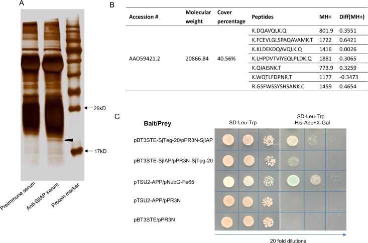
Schistosomes are causative agents of human schistosomiasis, which is endemic in tropical and subtropical areas of the world. Adult schistosomes can survive in their final hosts for several decades, and they have evolved various strategies to overcome the host immune response. Consequently, understanding the mechanisms that regulate parasitic cell survival will open avenues for developing novel strategies against schistosomiasis. Our previous study suggested that an inhibitor of apoptosis protein in Schistosoma japonicum (SjIAP) may play important roles in parasitic survival and development. Here, we demonstrated that SjIAP can negatively regulate cellular apoptosis in S. japonicum by suppressing caspase activity. Immunohistochemistry analysis indicated that SjIAP ubiquitously expressed within the worm body including the tegument. Silencing of SjIAP expression via small interfering RNA led to destruction of the tegument integrity in schistosomes. We further used co-immunoprecipitation to identify interaction partners of SjIAP and revealed the tegument protein SjTeg-20 as a putative interacting partner of SjIAP. The interaction between SjIAP and SjTeg-20 was confirmed by a yeast two-hybrid (Y2H) assay. Moreover, results of a TUNEL assay, RNA interference, scanning and transmission electron microscopy, caspase assays, transcript profiling, and protein localization of both interacting molecules provided first evidence for an essential role of SjIAP and SjTeg-20 to maintain the structural integrity of the tegument by negatively regulating apoptosis. Taken together, our findings suggest that the cooperative activities of SjIAP and SjTeg-20 belong to the strategic inventory of S. japonicum ensuring survival in the hostile environment within the vasculature of the final host.


Conflict of interest statement

The authors have declared that no competing interests exist.

Figures

Fig 1
Fig 1. Developmental expression and localization of SjIAP in S. japonicum.
(A) Expression of SjIAP at different stages of the S. japonicum life cycles. Eggs were isolated from liver of rabbits infected with S. japonicum cercariae, cercariae were collected from infected Oncomelania hupensis snails, and the parasites were collected from infected rabbits at 7, 14, 21, 28, and 35 days post-infection, respectively. The mRNA expression levels of SjIAP relative to SjNADH were analyzed by qRT-PCR. Data illustrate representative results and show the mean and standard errors derived from triplicate experiments. (B) Immunohistochemistry analysis of SjIAP in adult schistosomes using an antiserum against SjIAP [21]. Arrows indicate the schistosome tegument. Scale bars: 50 μm.
Fig 2
Fig 2. Effect of SjIAP silencing on caspase activity and worm survival.
(A) Incubation of adult worms with SjIAP siRNA-951 in vitro led to a significant decrease of SjIAP expression at the transcript level. At 4 days of post-treatment, the RNA was isolated from the worms. Transcript levels of SjIAP relative to the reference gene SjNADH were determined in parasites transfected with SjIAP siRNA and control siRNA using qRT-PCR. The data illustrate representative results and show mean±SEM derived from triplicate experiments. ** P ≤ 0.01. (B) Western blot analysis of the effect of SjIAP silencing at the protein level. The densitometry results (arbitrary units [AU]) of the Western blot analyzed by Image J were shown as a bar graph. Each bar represents mean±SEM from triplicate experiments. Representative Western blot result was also shown. ** P ≤ 0.01. (C) SjIAP silencing resulted in increased caspase 3/7 activity. At 4 days of post-treatment, the protein lysates of worms were prepared and used to determine caspase 3/7 activity. Data are representative result and shown the mean and standard errors from triplicate experiments. ** P ≤ 0.01. (D) qRT-PCR analysis of the transcript levels of different caspases (caspase 2, caspase 3, and caspase 7) in SjIAP-silenced and control schistosomes determined relative to SjNADH at 4 days post-treatment. (E) SjIAP silencing led to an increased number of TUNEL+ cells in the tegument (arrows) of schistosomes at 4 days post-treatment. Data are representative results from at least 20 worms investigated in at least three independent experiments. (F) SjIAP silencing resulted in increased worm mortality. At 4 days post-treatment, the worms were stained with Hoechst 33258 dye, and the mortality was determined by microscopy. Data are representative and show the mean and standard errors from three separate experiments. ** P ≤ 0.01.
Fig 3
Fig 3. SjIAP inhibition by siRNA led to morphological alterations in the tegument of S. japonicum.
(A) Scanning electronic microscopy (SEM) of the tegument of S. japonicum following treatment with SjIAP siRNA. Tegument ridges and sensory structures were observed in the mid part of the worm body. Data are representative results from at least 20 worms investigated in three independent experiments. (B) Transmission electronic microscopy (TEM) of the tegument of S. japonicum following treatment with SjIAP siRNA. Data are representative results from at least 15 worms investigated in three independent experiments. Scale bars: 2 μM. (C) Quantification of tegument changes due to SjIAP silencing shown in (B). Tegument defects were analyzed using Image J software based on the ratio of the area occupied by the tegument cytoplasm to the total area of the tegument. Data show the mean and standard errors derived from four randomly selected parasites. ** P ≤ 0.01 (Student’s t test, SjIAP siRNA vs control siRNA).
Fig 4
Fig 4. Identification and validation of SjTeg-20 as interaction partner of SjIAP.
(A) For co-immunoprecipitation, protein lysates from adult schistosomes were incubated with anti-SjIAP serum and pre-immune serum. The pull-down products were separated by SDS-PAGE and visualized by sliver staining. (B) Search results for MS identification of the co-immunoprecipitation products. (C) Validation of the interaction between SjIAP and SjTeg-20 by Y2H analysis. All diploid strains in which the density was equalized before plating grew equally well under selection for both the bait and prey plasmids (SD-Leu-Trp). Twenty-fold dilutions for each diploid were inoculated. Prey SjIAP and bait SjTeg-20 strongly interacted on the reporter plate (SD-Leu-Trp-Ade-His+ X-gal). Bait SjIAP and prey SjTeg-20 also interacted but weaker as the positive control (pTSU2-APP+pNubG-Fe65) on the reporter plate. No interaction was detected with the negative control and empty plasmids.
Fig 5
Fig 5. Expression profile of SjTeg-20 and its localization in S. japonicum.
(A) qRT-PCR analysis of the expression profiles of SjTeg-20 at different stages of S. japonicum relative to SjNADH. Data illustrate representative results and show the mean and standard errors derived from triplicate experiments. (B) Immunohistochemistry analysis of SjTeg-20 expression in the tegument (arrows) of adult schistosomes. Scale bars: 50 μm.
Fig 6
Fig 6. Effects of SjTeg-20 silencing on caspase activity, tegument destruction, and worm mortality.
(A) Screening of the best siRNA duplex for inhibiting SjTeg-20. Three siRNA duplexes were electroporated into in vitro cultured schistosomes, respectively, and SjTeg-20 mRNA levels were determined relative to SjNADH in SjTeg-20 siRNA and control siRNA-treated worms at 4 days post electroporation by qRT-PCR. Data illustrate representative results and show the mean and standard errors derived from triplicate experiments. ** P ≤ 0.01. (B) Validation of the SjTeg-20 siRNA-132 duplex for silencing SjTeg-20 using western blot analysis. The densitometry results (arbitrary units [AU]) of the Western blot analyzed by Image J were shown as a bar graph. Each bar represents mean±SEM from triplicate experiments. Representative Western blot result was also shown. ** P ≤ 0.01. (C) SjTeg-20 silencing resulted in morphological alterations in the tegument of S. japonicum as determined by SEM. Data are representative results from at least 15 worms investigated in three independent experiments. (D) SjTeg-20 silencing resulted in increased caspase 3/7 activity of cultured worms (28 d) in vitro which were electroporated with SjTeg-20 siRNA-132. (E) SjTeg-20 silencing increased worm mortality based on Hoechst 33258 dye staining and microscopic observations. Data are representative results and shown as the mean ± standard error from triplicate experiments. ** P ≤ 0.01 (Student’s t test, SjTeg-20 siRNA vs control siRNA).
Fig 7
Fig 7. SjIAP and SjTeg-20 synergistically regulate the apoptotic process and maintain the tegument architecture in S. japonicum.
(A) qRT-PCR and Western blot analysis to investigate the effect of SjTeg-20 silencing on the expression of SjIAP. qRT-PCR data are representative results and shown as the mean ± standard error from triplicate experiments. * P ≤ 0.05. (B) qRT-PCR and Western blot analysis of the effect of SjIAP silencing on the expression of SjTeg-20. qRT-PCR data are representative results and shown as the mean ± standard error from triplicate experiments. * P ≤ 0.05. (C) Co-silencing of SjIAP and SjTeg-20 significantly elevated caspase 3/7 activity determined from worm protein lysates. Data are representative results and shown as the mean ± standard error from triplicate experiments. ** P ≤ 0.01. (D) Effect of co-transfection of recombinant plasmids for expressing SjIAP and SjTeg-20 on the caspase activity in mammalian cells. HEK293T cells were treated with Cyclosporin A for 12 h and then transfected with recombinant plasmids expressing SjIAP and SjTeg-20 (2 μg). At 12 h post transfection, cells were collected for determining caspase 3/7 activity. Data are representative results and show the mean ± standard error from triplicate experiments. ** P ≤ 0.01. (E) Effect of co-transfection of the recombinant plasmids expressing SjIAP and SjTeg-20 on cell apoptosis in mammalian cells. HEK293T cells were treated with Cyclosporin A for 12 h, transfected with recombinant plasmids expressing SjIAP and SjTeg-20 (2 μg), stained with Annexin V-FITC kit, and subjected to flow cytometry. Data are representative results from triplicate experiments. (F) Quantitation of apoptosis cells transfected with recombinant plasmids expressing SjIAP and SjTeg-20 in HEK293T cells using flow cytometry. Data are representative result and shown the mean ± standard error from triplicate experiments. ** P ≤ 0.01. (G) TEM of morphological alterations in the tegument of S. japonicum treated with SjIAP siRNA and/or SjTeg-20 siRNA. Data are representative results from at least 15 worms investigated in three independent experiments. Scale bars: 2 μM. (H) Quantification of tegument changes due to co-silencing SjIAP and SjTeg-20 shown in (G). The tegument defects were analyzed using Image J based on the ratio of the area occupied by the tegument cytoplasm to the total area of the tegument. Data show the mean and standard errors derived from four randomly selected parasites. ** P ≤ 0.01. Abbreviations: Mus-muscles; teg-surface layer of the tegument. (I) Co-silencing SjIAP and SjTeg-20 resulted in significantly increased worm mortality. At 4 days of post treatment, worms were stained with Hoechst 33258 dye, and the mortality was determined by microscopy. Data are representative results and shown as the mean ± standard error from triplicate experiments. ** P ≤ 0.01.
Fig 8
Fig 8. Silencing of SjIAP and SjTeg-20 decreased worm burden and egg deposition in mice in vivo.
(A) Schematic showing the schedule for siRNA injection in mice. Each treatment included 6 mice (n = 6) (B) qRT-PCR analysis of SjIAP mRNA from surviving schistosomes isolated from mice injected with SjIAP siRNA by hydrodynamic tail vein injection, determined relative to SjNADH. Data are shown as the mean ± standard error and are representative results from triplicate experiments. ** P ≤ 0.01. (C) Effect of SjIAP siRNA injection on the worm burden in mice. At 14 days post infection, mice were administrated with SjIAP siRNA five times with one day interval. At 28 days of post infection, worms were perfused from mice, and the number of worms was counted. ** P ≤ 0.01. (D) Effect of SjIAP siRNA injection on egg deposition in the liver of mice. ** P ≤ 0.01. (E) qRT-PCR analysis of SjIAP mRNA levels from schistosomes isolated from mice injected with SjIAP/SjTeg-20 siRNA by hydrodynamic tail vein injection in the biological replicates. Levels of SjIAP mRNA relative to SjNADH were analyzed by qRT-PCR in surviving worms used for RNA isolation. Data are shown as the mean ± standard error and representative results from triplicate experiments. * P ≤ 0.05 and ** P ≤ 0.01. (F) qRT-PCR analysis of SjTeg-20 mRNA level from schistosomes isolated from mice injected with SjIAP/SjTeg-20 siRNAs by hydrodynamic tail vein injection in the biological replicates. Levels of SjTeg-20 mRNA relative to SjNADH were analyzed by qRT-PCR in surviving worms used for RNA isolation. Data are shown as mean ± standard error and representative results from triplicate experiments. ** P ≤ 0.01. (G) Effect of SjIAP/SjTeg-20 siRNA injection on the worm burden in mice. At 14 days post infection, mice were administered with SjIAP/SjTeg-20 siRNA as indicated above. At 28 days of post infection, worms were perfused from mice, and the number of worms was counted. * P ≤ 0.05 and ** P ≤ 0.01. (H) Effect of SjIAP/SjTeg-20 siRNA injection on egg deposition in the liver of mice. * P ≤ 0.05 and ** P ≤ 0.01.

References

    1. Hotez PJ, Alvarado M, Basanez MG, Bolliger I, Bourne R, Boussinesq M, et al. The global burden of disease study 2010: interpretation and implications for the neglected tropical diseases. PLoS Negl Trop Dis. 2014;8(7):e2865 10.1371/journal.pntd.0002865 - DOI - PMC - PubMed
    1. Bergquist R, Utzinger J, Keiser J. Controlling schistosomiasis with praziquantel: How much longer without a viable alternative? Infect Dis Poverty. 2017;6(1):74 10.1186/s40249-017-0286-2 - DOI - PMC - PubMed
    1. Kerr JF, Wyllie AH, Currie AR. Apoptosis: a basic biological phenomenon with wide-ranging implications in tissue kinetics. Br J Cancer. 1972;26(4):239–57. - PMC - PubMed
    1. Williams GT. Programmed cell death: a fundamental protective response to pathogens. Trends Microbiol. 1994;2(12):463–4. - PubMed
    1. Peng J, Gobert GN, Hong Y, Jiang W, Han H, McManus DP, et al. Apoptosis governs the elimination of Schistosoma japonicum from the non-permissive host Microtus fortis. PLoS One. 2011;6(6):e21109 10.1371/journal.pone.0021109 - DOI - PMC - PubMed

Publication types