

Glia-neuron energy metabolism in health and diseases: New insights into the role of nervous system metabolic transporters
- PMID: 30044944
- PMCID: PMC6156776
- DOI: 10.1016/j.expneurol.2018.07.009
Glia-neuron energy metabolism in health and diseases: New insights into the role of nervous system metabolic transporters
Abstract
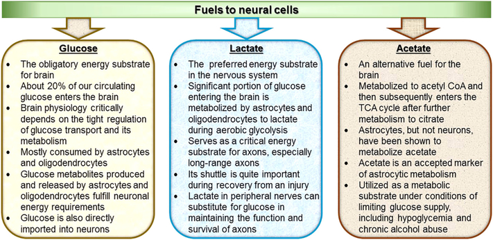
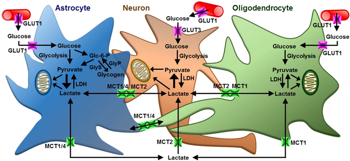
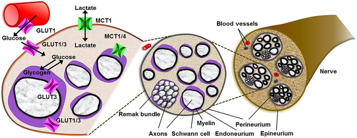
The brain is, by weight, only 2% the volume of the body and yet it consumes about 20% of the total glucose, suggesting that the energy requirements of the brain are high and that glucose is the primary energy source for the nervous system. Due to this dependence on glucose, brain physiology critically depends on the tight regulation of glucose transport and its metabolism. Glucose transporters ensure efficient glucose uptake by neural cells and contribute to the physiology and pathology of the nervous system. Despite this, a growing body of evidence demonstrates that for the maintenance of several neuronal functions, lactate, rather than glucose, is the preferred energy metabolite in the nervous system. Monocarboxylate transporters play a crucial role in providing metabolic support to axons by functioning as the principal transporters for lactate in the nervous system. Monocarboxylate transporters are also critical for axonal myelination and regeneration. Most importantly, recent studies have demonstrated the central role of glial cells in brain energy metabolism. A close and regulated metabolic conversation between neurons and both astrocytes and oligodendroglia in the central nervous system, or Schwann cells in the peripheral nervous system, has recently been shown to be an important determinant of the metabolism and function of the nervous system. This article reviews the current understanding of the long existing controversies regarding energy substrate and utilization in the nervous system and discusses the role of metabolic transporters in health and diseases of the nervous system.
Keywords: Acetate; Axon; Energy metabolism; Glia; Glucose; Glucose transporters; Lactate; Metabolic transporters; Monocarboxylate transporters.
Copyright © 2018 Elsevier Inc. All rights reserved.
Figures
References
-
- Allen A, Messier C, 2013. Plastic changes in the astrocyte GLUT1 glucose transporter and beta-tubulin microtubule protein following voluntary exercise in mice. Behav. Brain Res 240, 95–102. - PubMed
-
- Allt G, Lawrenson JG, 2000. The blood-nerve barrier: enzymes, transporters and receptors–a comparison with the blood-brain barrier. Brain Res. Bull 52 (1), 1–12. - PubMed
-
- Arluison M, Quignon M, Nguyen P, Thorens B, Leloup C, Penicaud L, 2004. Distribution and anatomical localization of the glucose transporter 2 (GLUT2) in the adult rat brain—an immunohistochemical study. J. Chem. Neuroanat 28 (3), 117–136. - PubMed
-
- Arluison M, Quignon M, Thorens B, Leloup C, Penicaud L, 2004. Immunocytochemical localization of the glucose transporter 2 (GLUT2) in the adult rat brain. II. Electron microscopic study. J. Chem. Neuroanat 28 (3), 137–146. - PubMed
Publication types
MeSH terms
Substances
Grants and funding
LinkOut - more resources
Full Text Sources
Other Literature Sources
Medical
