Comparative Study
. 2018 Nov;42(11):1456-1465.
doi: 10.1097/PAS.0000000000001134.

PRAME Expression in Melanocytic Tumors

Affiliations
Comparative Study

PRAME Expression in Melanocytic Tumors

Cecilia Lezcano et al. Am J Surg Pathol. 2018 Nov.

Abstract

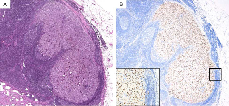
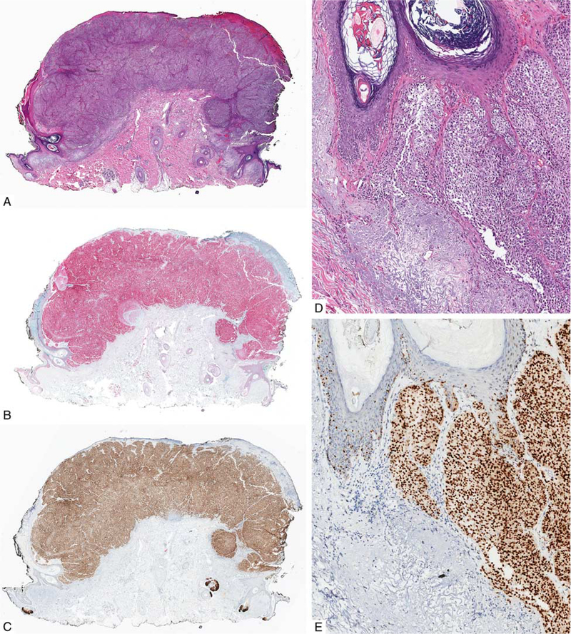
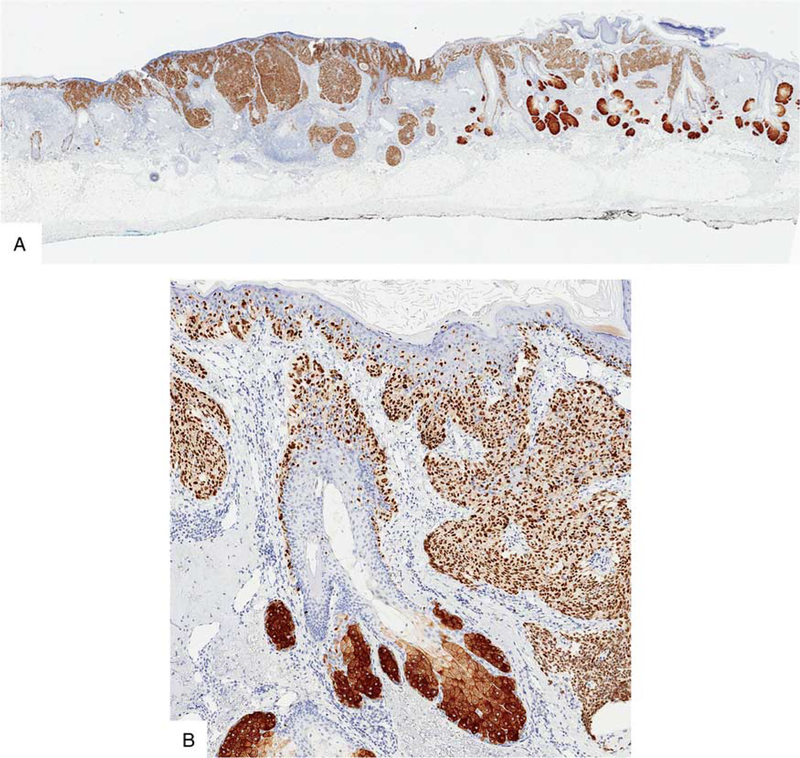
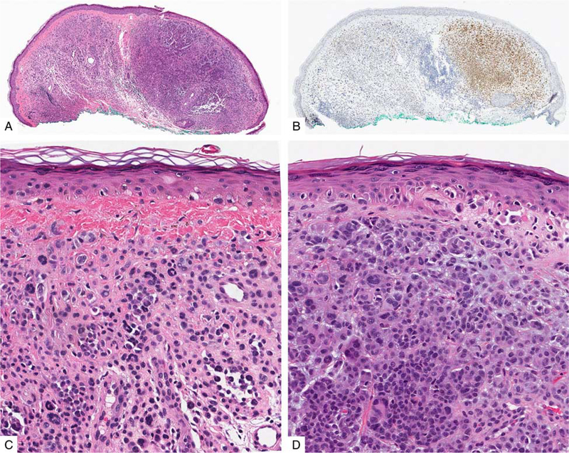
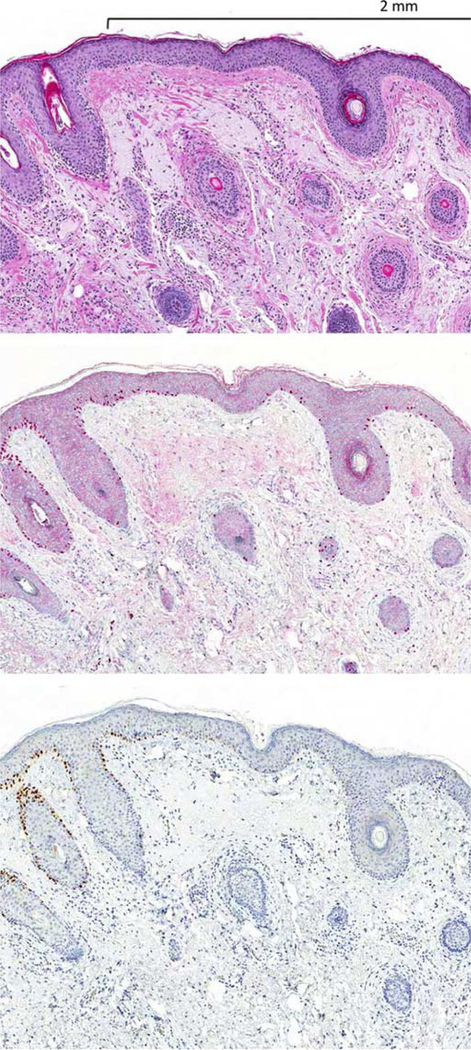
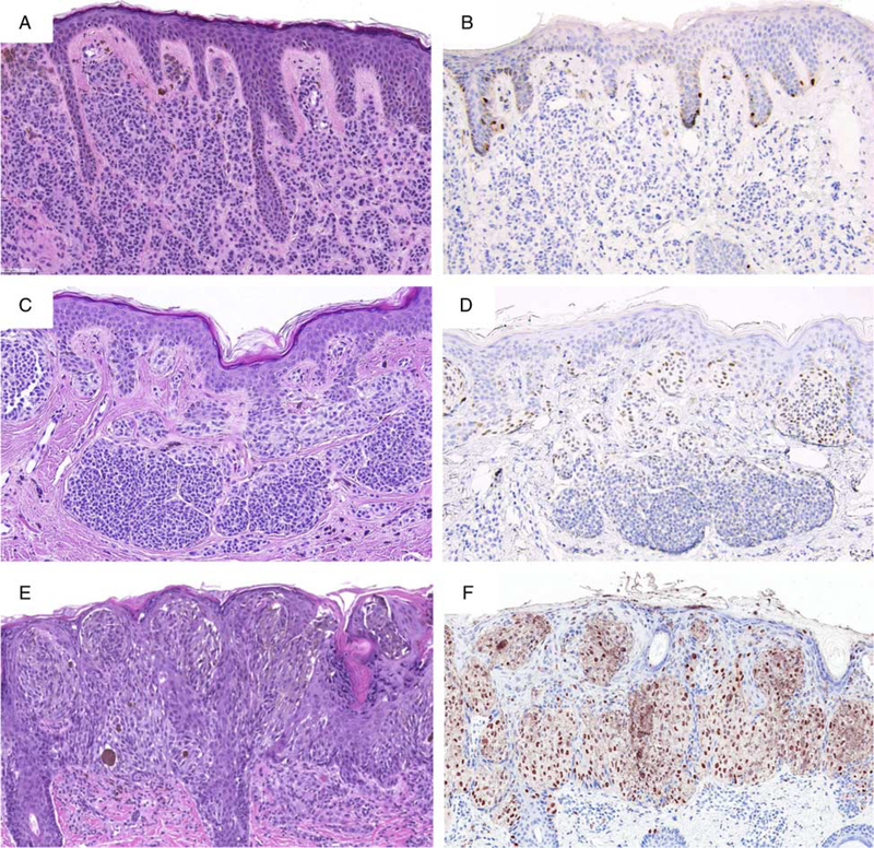
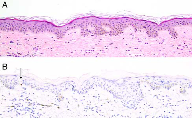
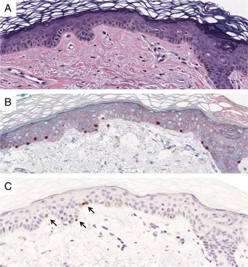
PRAME (PReferentially expressed Antigen in MElanoma) is a melanoma-associated antigen that was isolated by autologous T cells in a melanoma patient. While frequent PRAME mRNA expression is well documented in cutaneous and ocular melanomas, little is known about PRAME protein expression in melanocytic tumors. In this study we examined the immunohistochemical expression of PRAME in 400 melanocytic tumors, including 155 primary and 100 metastatic melanomas, and 145 melanocytic nevi. Diffuse nuclear immunoreactivity for PRAME was found in 87% of metastatic and 83.2% of primary melanomas. Among melanoma subtypes, PRAME was diffusely expressed in 94.4% of acral melanomas, 92.5% of superficial spreading melanomas, 90% of nodular melanomas, 88.6% of lentigo maligna melanomas, and 35% of desmoplastic melanomas. When in situ and nondesmoplastic invasive melanoma components were present, PRAME expression was seen in both. Of the 140 cutaneous melanocytic nevi, 86.4% were completely negative for PRAME. Immunoreactivity for PRAME was seen, albeit usually only in a minor subpopulation of lesional melanocytes, in 13.6% of cutaneous nevi, including dysplastic nevi, common acquired nevi, traumatized/recurrent nevi, and Spitz nevi. Rare isolated junctional melanocytes with immunoreactivity for PRAME were also seen in solar lentigines and benign nonlesional skin. Our results suggest that immunohistochemical analysis for PRAME expression may be useful for diagnostic purposes to support a suspected diagnosis of melanoma. It may also be valuable for margin assessment of a known PRAME-positive melanoma, but its expression in nevi, solar lentigines, and benign nonlesional skin can represent a pitfall and merits further investigations to better assess the potential clinical utility of this marker.


Figures

FIGURE 1.
FIGURE 1.
A, Metastatic melanoma in lymph node (H&E-stain). B, The tumor cells are diffusely immunopositive for PRAME (nuclear labeling). inset highlights PRAME labeling is nuclear.
FIGURE 2.
FIGURE 2.
Primary melanoma. A, Ulcerated polypoid tumor from the neck of a 73-year-old man. B, IHC for Sox10: the tumor cells are homogenously immunoreactive for Sox10. C, IHC for PRAME: the tumor cells diffusely express PRAME. D, Melanoma is present in both the epidermis and dermis (H&E-stained section). E, IHC for PRAME highlights both in situ and invasive tumor cells.
FIGURE 3.
FIGURE 3.
Primary melanoma from the scalp of a 75-year-old man. A, Both in situ and invasive melanoma are equally strongly immunoreactive for PRAME. There is prominent follicular involvement by melanoma. B, The melanocytes show nuclear labeling for PRAME. The sebaceous glands show cytoplasmic labeling.
FIGURE 4.
FIGURE 4.
Melanoma associated with a melanocytic nevus in the ear of a 63-year-old man. A, Nodular silhouette of the lesion with a more densely cellular tumor cell population on the right side of the lesion. B, IHC for PRAME stains only the densely cellular nodule. C, The less cellular area shows cytologic features of a melanocytic nevus. D, The PRAME-positive tumor cells are cytologically atypical. Cytogenetic analysis of the tumor cells revealed a number of chromosomal aberrations, including loss of 9p and gain of 8q (not shown).
FIGURE 5.
FIGURE 5.
Section from a staged excision of a lentigo maligna melanoma in situ with a rim of normal skin. A, H&E-stained section. The left blue-inked section edge faced the tumor debulk. The right side represents the outer rim—margin—of the excision. B, IHC for Sox10. C, IHC for PRAME. While the melanoma in situ strongly labels for PRAME, the melanocytes of the adjacent benign skin are negative.
FIGURE 6.
FIGURE 6.
PRAME immunoreactivity in nevi. A, Ordinary melanocytic nevus (H&E-stain). B, A few junctional melanocytes express PRAME (1+). C, Compound dysplastic nevus (H&E-stain). D, The center of the lesion contains a number of PRAME-positive melanocytes at the dermoepidermal junction and in the superficial dermis (2+). E, Predominantly junctional Spitz nevus on the cheek of a child (H&E-stain). F, The intraepidermal lesional melanocytes diffusely label for PRAME (4+).
FIGURE 7.
FIGURE 7.
A, Solar lentigo (H&E). B, A rare isolated junctional melanocyte labels for PRAME (arrow).
FIGURE 8.
FIGURE 8.
A, Benign sun-damaged skin (H&E). B, An immunostain for Sox10 documents cytologically bland benign junctional melanocytes. C, Rare junctional melanocytes show nuclear labeling for PRAME (arrows). Most melanocytes are negative for PRAME.

References

    1. Ikeda H, Lethe B, Lehmann F, et al. Characterization of an antigen that is recognized on a melanoma showing partial HLA loss by CTL expressing an NK inhibitory receptor. Immunity. 1997;6:199–208. - PubMed
    1. Hemminger JA, Toland AE, Scharschmidt TJ, et al. Expression of cancer-testis antigens MAGEA1, MAGEA3, ACRBP, PRAME, SSX2, and CTAG2 in myxoid and round cell liposarcoma. Mod Pathol. 2014;27:1238–1245. - PMC - PubMed
    1. Iura K, Kohashi K, Hotokebuchi Y, et al. Cancer-testis antigens PRAME and NY-ESO-1 correlate with tumour grade and poor prognosis in myxoid liposarcoma. J Pathol Clin Res. 2015;1:144–159. - PMC - PubMed
    1. Iura K, Maekawa A, Kohashi K, et al. Cancer-testis antigen expression in synovial sarcoma: NY-ESO-1, PRAME, MAGEA4, and MAGEA1. Hum Pathol. 2017;61:130–139. - PubMed
    1. Neumann E, Engelsberg A, Decker J, et al. Heterogeneous expression of the tumor-associated antigens RAGE-1, PRAME, and glycoprotein 75 in human renal cell carcinoma: candidates for T-cell-based immunotherapies? Cancer Res. 1998;58:4090–4095. - PubMed

Publication types

MeSH terms