. 2018 Sep 13;132(11):1134-1145.
doi: 10.1182/blood-2017-08-802926. Epub 2018 Jul 25.

Antitumor activity of CAR-T cells targeting the intracellular oncoprotein WT1 can be enhanced by vaccination

Affiliations

Antitumor activity of CAR-T cells targeting the intracellular oncoprotein WT1 can be enhanced by vaccination

Yasushi Akahori et al. Blood. .

Abstract

The recent success of chimeric antigen receptor (CAR)-T cell therapy for treatment of hematologic malignancies supports further development of treatments for both liquid and solid tumors. However, expansion of CAR-T cell therapy is limited by the availability of surface antigens specific for the tumor while sparing normal cells. There is a rich diversity of tumor antigens from intracellularly expressed proteins that current and conventional CAR-T cells are unable to target. Furthermore, adoptively transferred T cells often suffer from exhaustion and insufficient expansion, in part, because of the immunosuppressive mechanisms operating in tumor-bearing hosts. Therefore, it is necessary to develop means to further activate and expand those CAR-T cells in vivo. The Wilms tumor 1 (WT1) is an intracellular oncogenic transcription factor that is an attractive target for cancer immunotherapy because of its overexpression in a wide range of leukemias and solid tumors, and a low level of expression in normal adult tissues. In the present study, we developed CAR-T cells consisting of a single chain variable fragment (scFv) specific to the WT1235-243/HLA-A*2402 complex. The therapeutic efficacy of our CAR-T cells was demonstrated in a xenograft model, which was further enhanced by vaccination with dendritic cells (DCs) loaded with the corresponding antigen. This enhanced efficacy was mediated, at least partly, by the expansion and activation of CAR-T cells. CAR-T cells shown in the present study not only demonstrate the potential to expand the range of targets available to CAR-T cells, but also provide a proof of concept that efficacy of CAR-T cells targeting peptide/major histocompatibility complex can be boosted by vaccination.


Conflict of interest statement

Conflict-of-interest disclosure: Y. Amaishi, S. Okamoto, and J.M. are employees of Takara Bio Inc. which collaborated in the development of virus vector construction. The remaining authors declare no competing financial interests.

Figures

None
Graphical abstract
Figure 1.
Figure 1.
Design and characterization of a WT1235-243/HLA-A*2402-specific CAR. (A) Schematic representation of a retroviral vector encoding WT1235-243/HLA-A*2402-specific CAR. (B) Expression of CAR on human T cells. Human PBMCs stimulated with plate coated anti-CD3 were transduced with the retroviral vector as depicted in panel A. Four days after the transduction, cells were stained with phycoerythrin-labeled HLA-A*2402 tetramers presenting WT1236Y along with APC anti-CD4 and APC/Cy7 anti-CD8. Mock-transduced T cells served as background staining. (C) Production of IFN-γ by #213 scFv CAR-T cells stimulated with immobilized HLA-A*2402 tetramers loaded with WT1235-243 or WT1236Y. Culture supernatants were harvested at 24 hours and subjected to IFN-γ enzyme-linked immunosorbent assay in triplicate. (D) Cytotoxic activity of #213 scFv CAR-T cells against T2A24 cells pulsed with the indicated peptides at 10 μM was assayed by 6-hour 51Cr-release assays in triplicate. (E) IFN-γ production of #213 scFv CAR-T cells stimulated with T2A24 cells pulsed with the indicated peptides. % IFN-γ–positive cells within CD8+ cells of #213 scFv CAR-T cells were assayed by intracellular cytokine staining after a 6-hour culture in single experimental samples. Error bars represent standard deviation (SD) of the mean. A representative result of 3 independent experiments is shown.
Figure 2.
Figure 2.
#213 scFv CAR-T cells react to tumor cell lines endogenously expressing WT1 in an HLA-A*2402–restricted manner. (A) IFN-γ production of CD8+ cells of #213 scFv CAR-T cells in response to the indicated tumor cell lines was assayed by intracellular cytokine staining after a 6-hour culture (top) along with HLA-A*2402 (middle) and WT1 mRNA expression of those tumor cell lines (bottom). (B) Cytotoxic activity of #213 scFv CAR-T cells against the indicated tumor cell lines was assessed by 6-hour 51Cr-release assays in triplicate. (C) Confirmation of epitope specificity of #213 scFv CAR-T cells against peptide derived from endogenously expressing WT1. A standard 51Cr release assay using purified CD8+CAR+ T cells as effectors and a MESO-4 cell line as a target at an effector-to-target ratio of 2:1 was conducted in the presence of 40-fold excess of unlabeled T2A24 pulsed with either WT1236Y or MAGE-A4143-151. Error bars represent SD of the mean. A representative result of 3 independent experiments is shown.
Figure 3.
Figure 3.
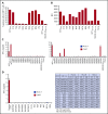
Potential cross-reactivity of #213 scFv CAR-T cells to homologous peptides on HLA-A*2402. (A) Alanine substitution analysis identified WT1235 peptide residues important for recognition by #213 scFv CAR. The WT1236Y peptide sequence was substituted with alanine from 1 through 9. T2A24 was pulsed with the indicated peptides at 10 μM and cocultured with #213 scFv CAR-T cells for 6 hours. IFN-γ–positive cells gated on CD8+ cells in CAR-T cells were assayed by intracellular cytokine staining. (B) The peptides in panel A were subjected to an HLA stabilization assay to determine the critical amino acids required for HLA-A*2402 binding. T2A24 cells were used as peptide-loading cells and their HLA-A*2402 expression was determined by fluorescence-activated cell sorter (FACS). (C) Validation of potential risk peptides derived from the human proteome. T2A24 cells were pulsed with the indicated peptides (sequences provided in supplemental Table 1) at 10 μM and cocultured with #213 scFv CAR-T cells (black column) or mock-transduced T cells (white column) for 24 hours. (D) Validation of potential cross-reactivity to allo-HLAs. LCLs expressing different HLAs were cocultured with #213 scFv CAR-T cells (black column) or mock-transduced T cells (white column) for 24 hours. T2A24 cells pulsed with 10 μM WT1236Y served as a positive control. Culture supernatants from these cultures were subjected to IFN-γ enzyme-linked immunosorbent assay in triplicate. Error bars represent SD of the mean. A representative result of 3 independent experiments is shown.
Figure 4.
Figure 4.
Adoptive transfer of WT1-specific CAR-T cells suppressed tumor growth in an antigen-specific manner. (A) Schematic representation for the adoptive transfer experiment using NOG mice. (B) Tumor growth curves of K562 and K562-A24 in NOG mice (n = 4) transferred with CAR-T cells or mock transduced T cells. NOG mice were inoculated s.c. with K562 and K562-A24 (5 × 10⁶ cells) followed by IV injection with #213 CAR-T cells or mock transduced T cells (1 × 10⁷ cells). Tumor volumes were measured by a caliper using the formula (length × width) at the indicated time points (n = 5). Error bars represent SD of the mean. *P < .05. A representative result from 2 independent experiments is shown. s.c., subcutaneous; TBI, total body irradiation.
Figure 5.
Figure 5.
Proliferation of #213 scFv CAR-T cells in response to WT1236Y directly presented by DCs. CFSE-labeled #213 scFv CAR-T cells were cocultured with HLA-A*2402+ DCs with WT1236Y peptide (WT1236Y SP) and subjected to CFSE-dilution assay on day 3. DCs pulsed with 10 μM MAGE-A4 peptides served as a negative control. A representative result from 3 independent experiments is shown.
Figure 6.
Figure 6.
Vaccination with DCs pulsed with WT1236Y enhanced WT1-specific CAR-T cells to suppress growth of WT1-positive tumors. (A) Schematic representation for the adoptive transfer experiment using NOG mice. (B) Tumor growth curves of K562-A24 in NOG mice (n = 4) transferred with CAR-T cells together with or without DCs pulsed with a relevant or irrelevant peptide. NOG mice bearing 3-day-old K562-A24 tumors were transferred with #213 scFv CAR-T cells or CEA-specific (F11-39 scFv) CAR-T cells (1 × 10⁷ cells) together with or without DCs (1 × 10⁵ cells) pulsed with WT1236Y or MAGE-A4143-151. Tumor volumes were measured by a caliper using the formula (length × width) at the indicated time points. Error bars represent SD of the mean. *P < .05; **P < .01. A representative result from 2 independent experiments is shown.
Figure 7.
Figure 7.
Antigen-specific DC vaccination enhanced #213 scFv CAR-T cells accumulation in periphery and activation in tumor tissues. Three-day K562-A24 bearing NOG mice were transferred with #213 scFv CAR-T cells or CEA-specific (F11-39 scFv) CAR-T cells (1 × 10⁷ cells) followed by vaccination with DCs (1 × 10⁵ cells) pulsed with WT1236Y or MAGE-A4143-151 as Figure 6. On day 16, PBMCs and tumor tissues were harvested and subjected to FACS (A) and immunohistochemical (B) analysis, respectively. A representative result from 2 independent experiments is shown.
Figure 8.
Figure 8.
Adoptive transfer with #213 scFv CAR-T cells suppressed growth of mesothelioma cell line, which was enhanced by the DC vaccine. Three-day MESO-1– or MESO-4–bearing NOG mice (n = 4) were transferred with #213 scFv CAR-T cells or CEA-specific (F11-39 scFv) CAR-T cells (1 × 10⁷ cells) followed by vaccination with DCs (1 × 10⁵ cells) pulsed with WT1236Y or MAGE-A4143-151. (A) Tumor volumes were measured by a caliper using the formula (length × width) at the indicated time points. (B) On day 24, PBMCs and tumor tissues were harvested and subjected to FACS analysis. In another set of experiments, tumor tissues were harvested from MESO-4 bearing NOG mice (n = 3) treated as mentioned previously on day 18 and subjected to immunohistochemical analysis. Cells with respective markers were analyzed in 6 fields, and the mean ± SD are depicted (C). Error bars represent SD of the mean. *P < .05; **P < .01. A representative result from 2 independent experiments is shown.

References

    1. Davila ML, Riviere I, Wang X, et al. . Efficacy and toxicity management of 19-28z CAR T cell therapy in B cell acute lymphoblastic leukemia. Sci Transl Med. 2014;6(224):224ra25. - PMC - PubMed
    1. Kochenderfer JN, Dudley ME, Kassim SH, et al. . Chemotherapy-refractory diffuse large B-cell lymphoma and indolent B-cell malignancies can be effectively treated with autologous T cells expressing an anti-CD19 chimeric antigen receptor. J Clin Oncol. 2015;33(6):540-549. - PMC - PubMed
    1. Maude SL, Frey N, Shaw PA, et al. . Chimeric antigen receptor T cells for sustained remissions in leukemia. N Engl J Med. 2014;371(16):1507-1517. - PMC - PubMed
    1. Kalos M, Levine BL, Porter DL, et al. . T cells with chimeric antigen receptors have potent antitumor effects and can establish memory in patients with advanced leukemia. Sci Transl Med. 2011;3(95):95ra73. - PMC - PubMed
    1. Lee DW, Kochenderfer JN, Stetler-Stevenson M, et al. . T cells expressing CD19 chimeric antigen receptors for acute lymphoblastic leukaemia in children and young adults: a phase 1 dose-escalation trial. Lancet. 2015;385(9967):517-528. - PMC - PubMed

Publication types