. 2018 Jul 26;3(14):e98202.
doi: 10.1172/jci.insight.98202.

Exogenous LRRK2G2019S induces parkinsonian-like pathology in a nonhuman primate

Affiliations

Exogenous LRRK2G2019S induces parkinsonian-like pathology in a nonhuman primate

Nadine Mestre-Francés et al. JCI Insight. .

Abstract

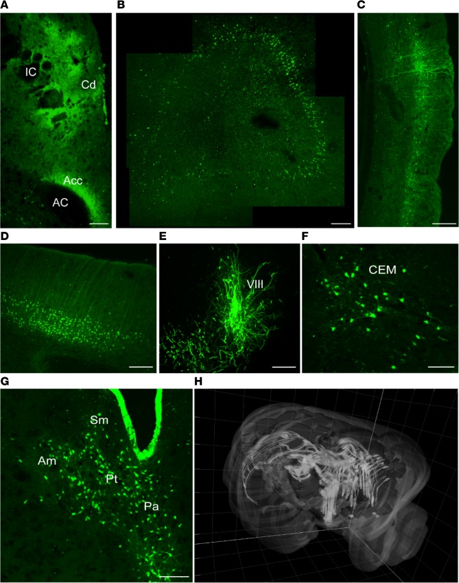
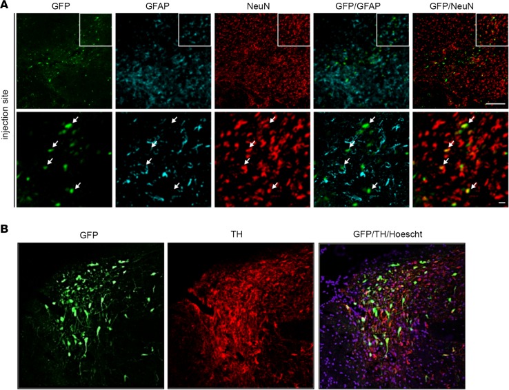
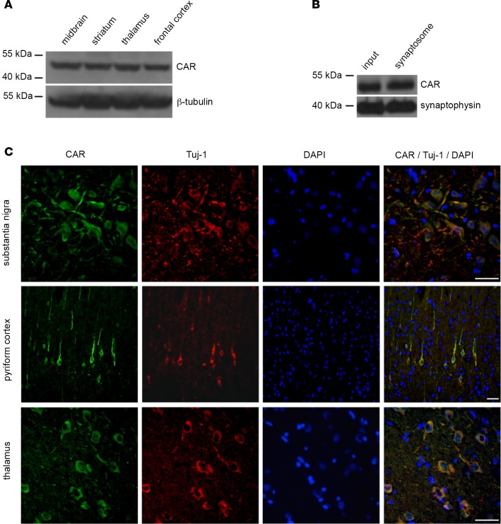
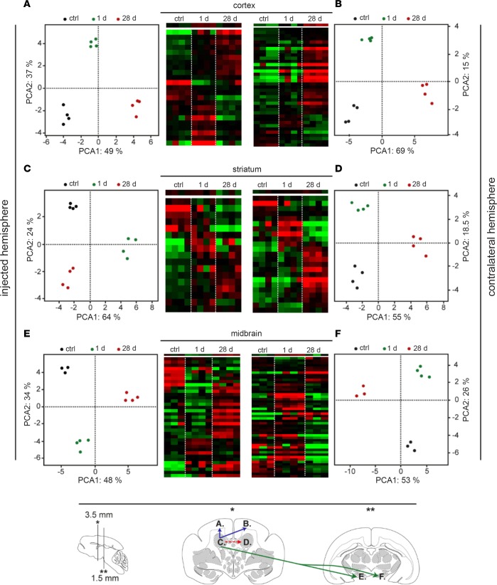
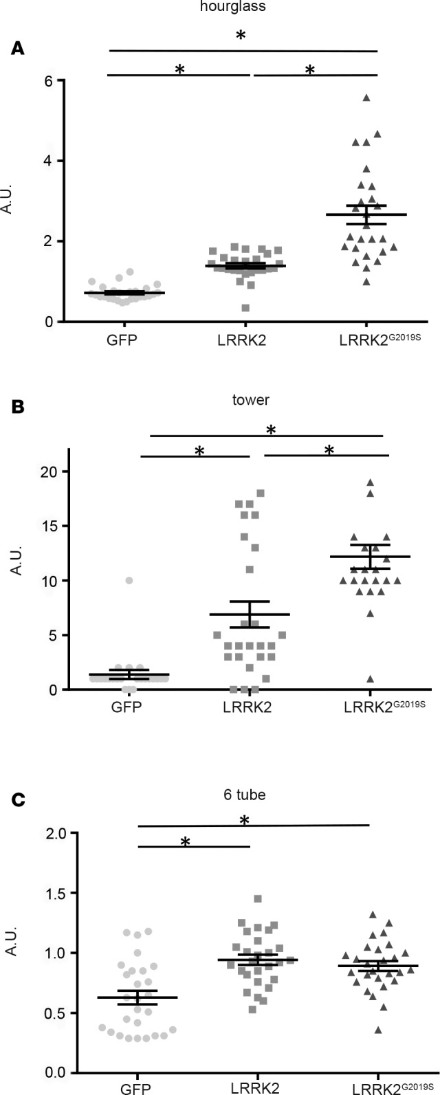
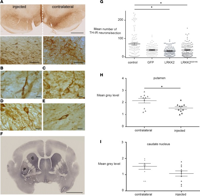
Parkinson's disease (PD) is the second most prevalent neurodegenerative disease among the elderly. To understand its pathogenesis and to test therapies, animal models that faithfully reproduce key pathological PD hallmarks are needed. As a prelude to developing a model of PD, we tested the tropism, efficacy, biodistribution, and transcriptional effect of canine adenovirus type 2 (CAV-2) vectors in the brain of Microcebus murinus, a nonhuman primate that naturally develops neurodegenerative lesions. We show that introducing helper-dependent (HD) CAV-2 vectors results in long-term, neuron-specific expression at the injection site and in afferent nuclei. Although HD CAV-2 vector injection induced a modest transcriptional response, no significant adaptive immune response was generated. We then generated and tested HD CAV-2 vectors expressing leucine-rich repeat kinase 2 (LRRK2) and LRRK2 carrying a G2019S mutation (LRRK2G2019S), which is linked to sporadic and familial autosomal dominant forms of PD. We show that HD-LRRK2G2019S expression induced parkinsonian-like motor symptoms and histological features in less than 4 months.

Keywords: Neurodegeneration; Neuroscience; Parkinson’s disease.


Conflict of interest statement

Conflict of interest: The authors have declared that no conflict of interest exists.

Figures

Figure 1
Figure 1. Expression of GFP following injection of HD-GFP in the M.
murinusstriatum. The brains from 6 M. murinus, injected at 8–33 months of age (see Table 1), were used to determine the efficacy of GFP expression from a helper-dependent (HD) CAV-2 vector (HD-GFP). (A) Injection site. Juxtaposed images showing the caudate nucleus (Cd), internal capsule (IC), nucleus accumbens (Acc), and anterior commissural (AC). (B) Ipsilateral prefrontal cortex (4 images juxtaposed). (C) Ipsilateral pyriform cortex. (D) Contralateral frontal cortex (cell bodies are located in the second and fourth cortical layers). (E) Substantia nigra (VIII). (F) Basal nucleus of Meynert (central-medial nucleus [CEM]). (G) Thalamic nuclei: periventricular nucleus (Pa), nucleus parataenialis (Pt), stria medullaris (Sm), anterior-medial nucleus (Am). (H) 3D reconstruction of the brain showing distribution of the GFP signal in both hemispheres following injection into the caudate nucleus. Images in A, C, F, and G are from M. murinus 133, which was sacrificed 6 months after injection. Scale bar: 200 μm.
Figure 2
Figure 2. Identification of HD-GFP–transduced cells in the M.
murinusbrain. Sections from M. murinus brains (n = 6 animals) were stained for neuronal and astrocytes specific markers to identify transduced cells. (A) Representative image from the injection site, caudate nucleus, showing GFP, glial fibrillary acidic protein (GFAP), and NeuN (neuronal nuclei antigen) expression followed by merged images. (B) Representative image from the substantia nigra (SN) showing GFP and tyrosine hydroxylase (TH) expression, followed by merged images. Scale bars: 100 μm (A, top) and 25 μm (A, bottom); and 100 μm (B).
Figure 3
Figure 3. Coxsackievirus and adenovirus receptor expression in the M.
murinusbrain. (A) Immunoblot analyses of coxsackievirus and adenovirus receptor (CAR) from the brains of M. murinus (n = 2 animals). β-Tubulin levels were used to normalize loading. (B) CAR levels in synaptosomes prepared from the cortex (n = 2 animals) and assayed for the presence of CAR. As a control, we used synaptophysin, a prototypic synapse protein. (C) CAR expression pattern, as assayed by immunofluorescence staining: SN, pyriform cortex, and thalamus (n = 3 animals). The 16-micron-thick floating sections were coincubated an anti-Tuj1 (β-III tubulin) antibody to identify neurons and with DAPI to label nuclei. Scale bar: 50 μm.
Figure 4
Figure 4. Transcriptional profiles of the most discriminant genes in the striatum, in the frontal lobe, and in the midbrain after HD-GFP injection.
Hierarchical clustering was obtained for each brain region: (A) right frontal cortex; (B) left frontal cortex; (C) injected (right) striatum; (D) contralateral striatum; (E) right midbrain; and (F) left midbrain. Each column corresponds to one animal, whereas each row corresponds to one gene (red: overexpressed genes; green: downregulated genes; black: genes without expression changes). Principal component analysis (PCA) of the 19–45 genes sorted by SAM or by ANOVA for each brain region analyzed. The x axis corresponds to the principal component 1 and the y axis to the principal component 2 (expressed in percentages). Projection of the individuals shows that each group of animal can be distinguished from the other irrespective of the brain structure. At the bottom of the figure, there is a schematic representation of the M. Murinus brain, indicating vector transport from the injected site, the right striatum, to the contralateral side (red hatched arrow), to the frontal cortex (blue arrow), and to the midbrain (green arrow). Brains were collected at 1 or 24 days after injection.
Figure 5
Figure 5. Behavior analyses following GFP, LRRK2, or LRRK2G2019S expression.
(A) Hourglass test (4 GFP-, 4 LRRK2-, and 8 LRRK2G2019S-expressing animals). (B) Tower task (6 GFP-, 4 LRRK2-, and 8 LRRK2G2019S-expressing animals). (C) Six-tube task (2 GFP-, 4 LRRK2-, and 8 LRRK2G2019S-expressing animals). Data are expressed as mean ± SEM. Nonparametric Kruskal-Wallis test: *P < 0.05. AU, arbitrary units; LRRK2, leucine-rich repeat kinase 2.
Figure 6
Figure 6. Changes in TH cell characteristics following LRRK2G2019S expression.
Representative images of (A) tyrosine hydroxylase (TH) immunoreactivity (dark brown staining) in a coronal section of the midbrain of LRRK2G2019S-expressing M. murinus (n = 8 animals). High-magnification images of TH+ cell bodies and projections in the hemisphere expressing LRRK2G2019S (left) versus the contralateral hemisphere (right) are shown below. Scale bars: 1 mm (top); 50 μm (bottom). (B) Swollen neurites and (C) dystrophic neurons due to LRRK2G2019S expression. (D) Neurons in control animals (n = 2). (E) Loss of neurites and (F) TH immunoreactivity due to LRRK2G2019S expression. Scale bars: 2.5 mm. (G) Quantification of TH+ cells in the substantia nigra of the LRRK2G2019S-expressing hemisphere versus controls (n = 3 mock-, 5 HD-GFP–, 7 HD-LRRK2G2019S–, and 4 HD-LRRK2-injected animals). TH+ neurons were quantified by counting cells in 1 of every 6 sections. The y axis represents the average number of TH+ neurons/section in all groups. Mann-Whitney test: *P < 0.05. (H) optical density of TH immunoreactivity in the putamen (n = 4 animals). (I) Optical density of TH immunoreactivity in the caudate nucleus (n = 3 animals). Data in H and I are expressed as mean ± SEM. Nonparametric Kruskal-Wallis test: *P < 0.05. C, caudate nucleus; P, putamen.

References

    1. Surmeier DJ, Obeso JA, Halliday GM. Selective neuronal vulnerability in Parkinson disease. Nat Rev Neurosci. 2017;18(2):101–113. - PMC - PubMed
    1. Gloeckner CJ, et al. The Parkinson disease causing LRRK2 mutation I2020T is associated with increased kinase activity. Hum Mol Genet. 2006;15(2):223–232. doi: 10.1093/hmg/ddi439. - DOI - PubMed
    1. Steger M, et al. Phosphoproteomics reveals that Parkinson’s disease kinase LRRK2 regulates a subset of Rab GTPases. Elife. 2016;5:e12813. - PMC - PubMed
    1. Healy DG, et al. Phenotype, genotype, and worldwide genetic penetrance of LRRK2-associated Parkinson’s disease: a case-control study. Lancet Neurol. 2008;7(7):583–590. doi: 10.1016/S1474-4422(08)70117-0. - DOI - PMC - PubMed
    1. Guo L, Gandhi PN, Wang W, Petersen RB, Wilson-Delfosse AL, Chen SG. The Parkinson’s disease-associated protein, leucine-rich repeat kinase 2 (LRRK2), is an authentic GTPase that stimulates kinase activity. Exp Cell Res. 2007;313(16):3658–3670. doi: 10.1016/j.yexcr.2007.07.007. - DOI - PMC - PubMed

Publication types

MeSH terms

Substances