. 2018 Jul 25;9(1):2909.
doi: 10.1038/s41467-018-05244-y.

Soluble RANKL contributes to osteoclast formation in adult mice but not ovariectomy-induced bone loss

Affiliations

Soluble RANKL contributes to osteoclast formation in adult mice but not ovariectomy-induced bone loss

Jinhu Xiong et al. Nat Commun. .

Abstract

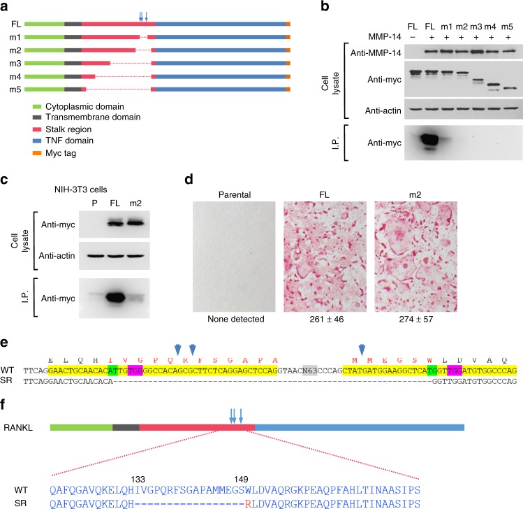
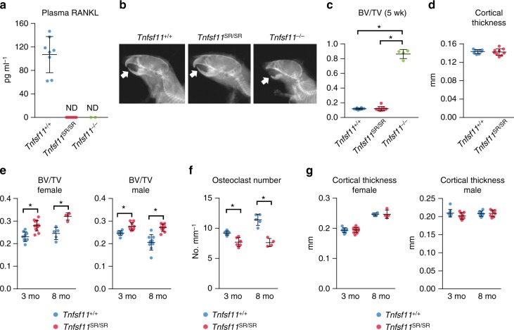
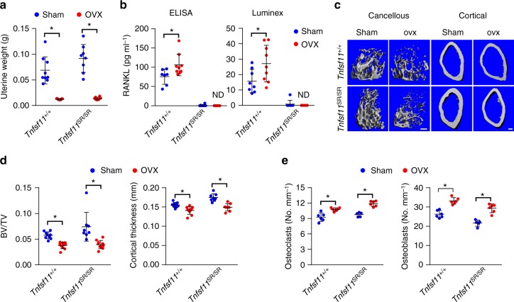
Receptor activator of NFkB ligand (RANKL) is a TNF-family cytokine required for osteoclast formation, as well as immune cell and mammary gland development. It is produced as a membrane-bound protein that can be shed to form a soluble protein. We created mice harboring a sheddase-resistant form of RANKL, in which soluble RANKL is undetectable in the circulation. Lack of soluble RANKL does not affect bone mass or structure in growing mice but reduces osteoclast number and increases cancellous bone mass in adult mice. Nonetheless, the bone loss caused by estrogen deficiency is unaffected by the lack of soluble RANKL. Lymphocyte number, lymph node development, and mammary gland development are also unaffected by the absence of soluble RANKL. These results demonstrate that the membrane-bound form of RANKL is sufficient for most functions of this protein but that the soluble form does contribute to physiological bone remodeling in adult mice.


Conflict of interest statement

C.A.O. owns stock in Radius Health, Inc. The remaining authors declare no competing interests.

Figures

Fig. 1
Fig. 1
Development of a sheddase-resistant RANKL. a Diagram of full-length (FL) and mutant RANKL constructs. Known cleavage sites are indicated by vertical arrows. b Sheddase assay in transiently transfected 293T cells. This experiment was replicated at least once. c Sheddase assay in stably transduced NIH-3T3 cells. P parental NIH-3T3 cells. This experiment was replicated at least once. d Osteoclast formation in co-cultures of bone marrow macrophages with the indicated NIH-3T3 cells shown in “c”. Osteoclasts are stained red. Quantification of multinucleated osteoclasts from triplicate wells shown below, mean ± s.d. This experiment was replicated at least once. e Partial DNA sequence of murine Tnfsf11 gene showing CRISPR/Cas9 gene-editing strategy. Exon 3 and a portion of exon 4 are highlighted in yellow. The sequence of wild-type (WT) and sheddase-resistant (SR) mutants are shown. N63 indicates 63 bp of intron 3 not shown. The PAM sequences for each of the two single-guide RNA (sgRNA) targets are highlighted in pink and their respective cut sites highlighted in green. Amino acids encoded by the exons are shown above the DNA sequence with the locations of known proteolytic cleavage sites indicated by the vertical blue arrows. The SR mutant was created by non-homologous end-joining of the double-stranded DNA cuts directed by the sgRNAs. f Diagram of a portion of the WT and SR RANKL proteins highlighting the amino acid sequences in the cleavage region. The red R indicates the position of an arginine resulting from deletion of the sequence shown in “e
Fig. 2
Fig. 2
Soluble RANKL contributes to osteoclast formation in adult mice. a sRANKL levels in the blood plasma of 5-week-old Tnfsf11+/+ (n = 8), Tnfsf11SR/SR (n = 9), and Tnfsr11−/− (n = 2) mice. ND not detectable. b X-ray images from 5-week-old Tnfsf11+/+, Tnfsf11SR/SR, and Tnfsr11−/− mice. Arrowheads indicate position of lower incisors. c Cancellous bone volume of L4 vertebra from 5-week-old Tnfsf11+/+ (n = 8), Tnfsf11SR/SR (n = 9), and Tnfsr11−/− (n = 4) mice. *P < 0.05 versus Tnfsf11+/+ or Tnfsf11SR/SR mice using one-way ANOVA. d Cortical thickness of femurs from 5-week-old Tnfsf11+/+ (n = 8) and Tnfsf11SR/SR (n = 9) mice. e Cancellous bone volume of L4 vertebra from 3-month-old female Tnfsf11+/+ (n = 9) and Tnfsf11SR/SR (n = 13) and male Tnfsf11+/+ (n = 8) and Tnfsf11SR/SR (n = 9) mice and 8-month-old female Tnfsf11+/+ (n = 5) and Tnfsf11SR/SR (n = 4) and male Tnfsf11+/+ (n = 9) and Tnfsf11SR/SR (n = 10) mice. *P < 0.05 versus Tnfsf11+/+ mice using Student’s t test. f Osteoclast number per mm bone surface measured in the cancellous bone of L1–L3 vertebra of 3-month-old Tnfsf11+/+ (n = 6) and Tnfsf11SR/SR (n = 6), and 8-month-old Tnfsf11+/+ (n = 5) and Tnfsf11SR/SR (n = 4), female littermates. *P < 0.05 using Student’s t test. g Cortical thickness of femurs from 3-month-old female Tnfsf11+/+ (n = 9) and Tnfsf11SR/SR (n = 13) and male Tnfsf11+/+ (n = 9) and Tnfsf11SR/SR (n = 9) mice and 8-month-old female Tnfsf11+/+ (n = 5) and Tnfsf11SR/SR (n = 4) and male Tnfsf11+/+ (n = 9) and Tnfsf11SR/SR (n = 10) mice. All analyses of 5-week-old mice included both sexes. All values are means ± s.d.
Fig. 3
Fig. 3
Soluble RANKL is not required for the bone loss caused by estrogen deficiency. a Uterine weight from Tnfsf11+/+ (sham n = 9, ovx n = 10) and Tnfsf11SR/SR (sham n = 8, ovx n = 8) mice. b Soluble RANKL in serum from Tnfsf11+/+ (sham n = 7, ovx n = 9) and Tnfsf11SR/SR (sham n = 8, ovx n = 7) mice measured by ELISA (left) or from Tnfsf11+/+ (sham n = 8, ovx n = 9) and Tnfsf11SR/SR (sham n = 8, ovx n = 8) mice measured by Luminex assay (right). c µCT images of femoral cancellous and cortical bone. Scale bar = 200 μm. d Cancellous bone volume (left) of femurs from Tnfsf11+/+ (sham n = 9, ovx n = 10) and Tnfsf11SR/SR (sham n = 8, ovx n = 10) mice and cortical thickness (right) of femurs from Tnfsf11+/+ (sham n = 9, ovx n = 10) and Tnfsf11SR/SR (sham n = 8, ovx n = 8) mice. e Osteoclast number (left) and osteoblast number (right) per mm bone surface measured in vertebral cancellous bone from Tnfsf11+/+ (sham n = 6, ovx n = 6) and Tnfsf11SR/SR (sham n = 5, ovx n = 6) mice. All values in Fig. 3 are from female mice sham-operated or ovariectomized at 3 months of age and analyzed 6 weeks later. All values are means ± s.d. *P < 0.05 versus sham-operated controls of the same genotype by Student’s t test

References

    1. Kong YY, et al. OPGL is a key regulator of osteoclastogenesis, lymphocyte development and lymph-node organogenesis. Nature. 1999;397:315–323. doi: 10.1038/16852. - DOI - PubMed
    1. Fujiwara Y, et al. RANKL (Receptor Activator of NFkappaB Ligand) produced by osteocytes is required for the increase in B cells and bone loss caused by estrogen deficiency in mice. J. Biol. Chem. 2016;291:24838–24850. doi: 10.1074/jbc.M116.742452. - DOI - PMC - PubMed
    1. Xiong J, et al. Matrix-embedded cells control osteoclast formation. Nat. Med. 2011;17:1235–1241. doi: 10.1038/nm.2448. - DOI - PMC - PubMed
    1. Fata JE, et al. The osteoclast differentiation factor osteoprotegerin-ligand is essential for mammary gland development. Cell. 2000;103:41–50. doi: 10.1016/S0092-8674(00)00103-3. - DOI - PubMed
    1. Knoop KA, et al. RANKL is necessary and sufficient to initiate development of antigen-sampling M cells in the intestinal epithelium. J. Immunol. 2009;183:5738–5747. doi: 10.4049/jimmunol.0901563. - DOI - PMC - PubMed

Publication types

LinkOut - more resources