

Diagnostic tools in the differential diagnosis of giant cell-rich lesions of bone at biopsy
- PMID: 30046391
- PMCID: PMC6059026
- DOI: 10.18632/oncotarget.25725
Diagnostic tools in the differential diagnosis of giant cell-rich lesions of bone at biopsy
Abstract
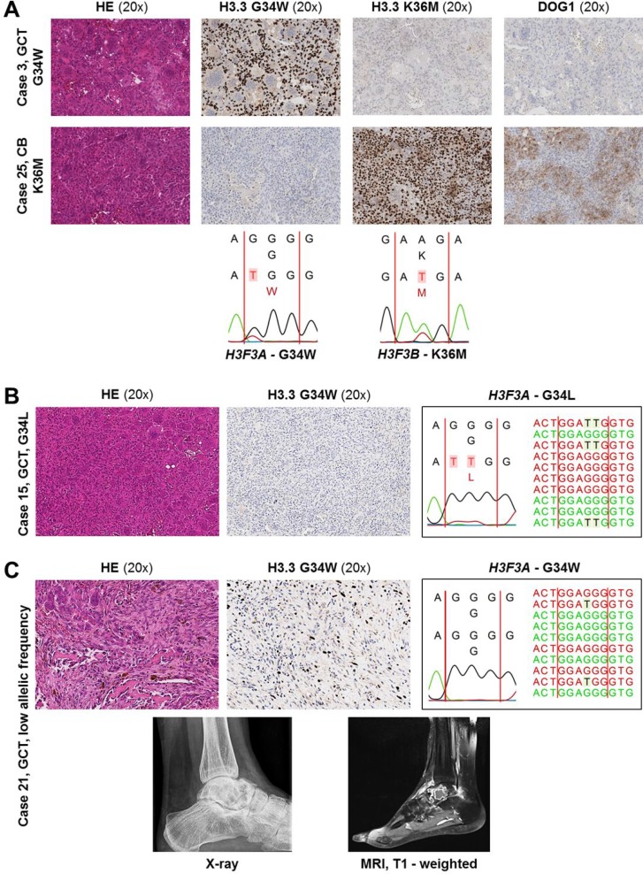
The diagnosis of giant cell-rich lesions of bone can be challenging if radiological findings are ambiguous and tissue of the biologically deciding component is underrepresented in biopsy specimens. The frequent association of giant cell tumor of bone (GCT) and chondroblastoma (CB) with (secondary) aneurysmal bone cysts (ABC) may further impede correct classification. The present study evaluates the potentials and limitations of mutation-specific histone H3.3 and DOG1 immunohistochemistry, Sanger-/next generation sequencing (NGS) and FISH analysis in the differential diagnosis of 23 GCT, 14 CB and 19 ABC. All morphologically typical GCT and CB harbored mutations in the H3F3A or H3F3B gene, respectively. These were, except for one uncommon G34L mutation in a GCT, reliably and specifically detected by mutation-specific H3.3 G34W or H3.3 K36M immunohistochemistry and DNA sequencing. In the diagnostic substantiation of CB, DOG1 staining was less sensitive compared to H3.3 K36M immunohistochemistry. 47% of ABC specifically showed translocations of the USP6 gene, while mutations in H3F3A/B were absent. Based on the results of this study, we conclude that mutation-specific H3.3 immunohistochemistry (selectively complemented with NGS-based DNA sequencing) and USP6 FISH analysis enable a reliable diagnostic distinction of GCT, CB and ABC of morphologically and radiologically difficult cases.
Keywords: H3F3A/B; aneurysmal bone cyst; chondroblastoma; diagnostic algorithm; giant cell tumor of bone.
Conflict of interest statement
CONFLICTS OF INTEREST The authors declare no conflicts of interest.
Figures


References
-
- Fletcher CD, Bridge JA, Hogendoorn PC, Mertens FE. WHO Classification of Tumours of Soft Tissue and Bone: International Agency for Research on Cancer. IARC. 2013
-
- Oliveira AM, Chou MM. USP6-induced neoplasms: the biologic spectrum of aneurysmal bone cyst and nodular fasciitis. Hum Pathol. 2014;45:1–11. - PubMed
-
- Atalar H, Basarir K, Yildiz Y, Erekul S, Saglik Y. Management of chondroblastoma: retrospective review of 28 patients. J Orthop Sci. 2007;12:334–340. - PubMed
-
- Levy WM, Miller AS, Bonakdarpour A, Aegerter E. Aneurysmal bone cyst secondary to other osseous lesions. Report of 57 cases. Am J Clin Pathol. 1975;63:1–8. - PubMed
-
- Dahlin DC, Ivins JC. Benign chondroblastoma. A study of 125 cases. Cancer. 1972;30:401–413. - PubMed
LinkOut - more resources
Full Text Sources
Other Literature Sources
