. 2018 Jun 26:2018:8481243.
doi: 10.1155/2018/8481243. eCollection 2018.

Positive Effects against UV-A Induced Damage and Oxidative Stress on an In Vitro Cell Model Using a Hyaluronic Acid Based Formulation Containing Amino Acids, Vitamins, and Minerals

Affiliations

Positive Effects against UV-A Induced Damage and Oxidative Stress on an In Vitro Cell Model Using a Hyaluronic Acid Based Formulation Containing Amino Acids, Vitamins, and Minerals

Antonietta Stellavato et al. Biomed Res Int. .

Retraction in

Abstract

Ultraviolet (UV) radiations are responsible for skin photoaging inducing alteration of the molecular and cellular pathways resulting in dryness and reduction of skin elasticity. In this study, we investigated, in vitro, the antiaging and antioxidant effects of hyaluronan formulations based hydrogel. Skinkò E, an intradermic formulation composed of hyaluronic acid (HA), minerals, amino acids, and vitamins, was compared with the sole HA of the same size. For this purpose, HaCaT cells were subjected to UV-A radiations and H2O2 exposure and then treated with growth medium (CTR) combined with M-HA or Skinkò E to evaluate their protective ability against stressful conditions. Cells reparation was evaluated using a scratch in vitro model and Time-Lapse Video Microscopy. A significant protective effect for Skinkò E was shown with respect to M-HA. In addition, Skinkò E increased cell reparation. Therefore, NF-kB, SOD-2, and HO-1 were significantly reduced at the transcriptional and protein level. Interestingly, γ-H2AX and protein damage assay confirmed the protection by hyaluronans tested against oxidative stress. G6pdΔ ES cell line, highly susceptible to oxidative stress, was used as a further cellular model to assess the antioxidant effect of Skinkò E. Western blotting analyses showed that the treatment with this new formulation exerts marked antioxidant action in cells exposed to UV-A and H2O2. Thus, the protective and reparative properties of Skinkò E make it an interesting tool to treat skin aging.


Figures

Figure 1
Figure 1
Hydrodynamic characterization by SEC-TDA is reported for (a) Skinkò E UF retentate and (b) M-HA.
Figure 2
Figure 2
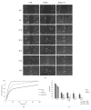
(a) Representative micrograph pictures of HaCaT scratch assays. (b) Repair area percentage for the control and in the presence of treatments: M-HA and Skinkò E; the curves are averages of three different experiments with standard deviation within 5% of the value. (c) Rate of reparation within 96 hours. Skinkò E and M-HA treatments are statistically significant  (p<0.05) with respect to CTR, Skinkò E and M-HA treatments are statistically significant  ∗∗(p<0.01) with respect to CTR, and Skinkò E is statistically significant §(p<0.05) with respect to M-HA treatment.
Figure 3
Figure 3
Antioxidant biomarkers (SOD-2 and HO-1) were determined by quantitative real-time PCR on HaCaT UV-A pretreated for 4 minutes and subsequently treated with M-HA 0.32% (w/v) and Skinkò E 0.32% (v/v) for 4 hours. Each column represents the mean and error bars represent the standard deviation (n=3). Test-t was performed to evaluate the significance of M-HA and Skinkò E effects with respect to positive control (UV-A),  p<0.01.
Figure 4
Figure 4
Western blotting analyses showed that M-HA and Skinkò E reduce the inflammatory proteins NF-kB and HO-1. In particular, Skinkò E is more efficient than M-HA on these protein modulations.
Figure 5
Figure 5
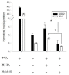
Effect of M-HA 0.32% (w/v) and Skinkò E 0.32% (v/v) on lipid peroxidation after exposure to oxidative stress induced by 50µM H2O2 for 30 minutes. M-HA and Skinkò E treatments are statistically significant (p<0.01) comparing with the positive control (H2O2). Also, Skinkò E is statistically significant (p<0.01) with respect to M-HA treatment.
Figure 6
Figure 6
Effect of M-HA 0.32% (w/v), Skinkò E 0.32% (v/v), and permeate (3KDa) Skinkò E on lipid peroxidation after exposure to oxidative stress induced by 50µM H2O2 (a) or UV-A irradiation (b) on HaCaT cells. “∗” in the histogram indicates significant difference at p<0.01, comparing with the Skinkò E and UF Skinkò E Permeate 3kDa treatment groups with respect to M-HA. Data of the amount of lipid peroxides (% respect to CTR) are reported as follows: Graph (a): H2O2=570±14, M-HA=128±7.0, Skinkò E=95.5±7.7, UF Skinkò E Permeate 3kDa=109±4.2. Graph (b): UV-A=480±14.0, M-HA=114±7.0, Skinkò E=74±5.6, UF Skinkò E Permeate 3kDa=76±7.0.
Figure 7
Figure 7
Protective effect of Skinkò E against oxidative stress-induced apoptosis.

References

    1. Gebbers N., Hurt-Burri N., Scaletta C., Hoffmann G., Applegate L. Water-filtered infrared-A radiation (wIRA) is not implicated in cellular degeneration of human skin, Ger Med Sci. Doc08: 5; 2007. - PMC - PubMed
    1. Thiesen L. C., Baccarin T., Fischer-Muller A. F., et al. Photochemoprotective effects against UVA and UVB irradiation and photosafety assessment of Litchi chinensis leaves extract. Journal of Photochemistry and Photobiology B: Biology. 2017;167:200–207. doi: 10.1016/j.jphotobiol.2016.12.033. - DOI - PubMed
    1. Svobodova A., Psotova J., Walterova D. Natural phenolics in the prevention of UV-induced skin damage: a review. Biomedical Papers. 2003;147:137–145. doi: 10.5507/bp.2003.019. - DOI - PubMed
    1. Yaar M., Gilchrest B. A. Photoageing: mechanism, prevention and therapy. British Journal of Dermatology. 2007;157(5):874–887. doi: 10.1111/j.1365-2133.2007.08108.x. - DOI - PubMed
    1. Xing Y.-X., Li P., Miao Y.-X., Du W., Wang C.-B. Involvement of ROS/ASMase/JNK signalling pathway in inhibiting UVA-induced apoptosis of HaCaT cells by polypeptide from Chlamys farreri. Free Radical Research. 2008;42(1):12–19. doi: 10.1080/10715760701762415. - DOI - PubMed

Publication types

LinkOut - more resources