. 2018 Jul 20;4(4):e248.
doi: 10.1212/NXG.0000000000000248. eCollection 2018 Aug.

Atypical Alexander disease with dystonia, retinopathy, and a brain mass mimicking astrocytoma

Affiliations

Atypical Alexander disease with dystonia, retinopathy, and a brain mass mimicking astrocytoma

Keren Machol et al. Neurol Genet. .
No abstract available


Figures

Figure
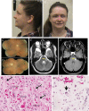
Figure. Clinical, histologic, and imaging findings
A 35-year-old woman with juvenile-onset Alexander disease (AD) (A and B). Note relative macrocephaly (FOC 56 cm, 94th centile) and esotropia. The photomontage of the right (C) and left (D) eyes shows the atrophic gliosis from prior optic disc edema and the marked retinal atrophy and secondary pigmentary degeneration. The retinal vasculature appears to show glial sheathing and peripheral vaso-obliteration in each eye. Brain MRI with contrast at 29 years (E and F). (E) Axial T2 image demonstrates a T2 hyperintense lesion centered in the left middle cerebellar peduncle (yellow arrow). (F) Axial T1 image with contrast shows an area of central enhancement within the lesion (yellow arrow). Partially imaged is cystic encephalomalacia within the frontal lobes bilaterally (yellow asterisk) with surgical changes in the right frontal lobe not included. Slides from patient's brain lesion (G and H) demonstrating AD histologic features. The prototypical histologic feature of AD is prominent Rosenthal fiber formation (G, solid arrows). An additional feature unique to AD is the presence of Rosenthal fiber-like eosinophilic cytoplasmic inclusions in astrocyte cell bodies (G, open arrows). The cell body inclusions are generally not seen in other Rosenthal fiber–rich conditions such as low-grade primary brain tumors. Astrocytes with markedly atypical nuclei (H, solid arrow) are also a characteristic morphologic feature of AD and can mimic the neoplastic cells of astrocytoma or ganglioglioma. FOC = fronto-occipital circumference.

References

    1. Prust M, Wang J, Morizono H, et al. . GFAP mutations, age at onset, and clinical subtypes in Alexander disease. Neurology 2011;77:1287–1294. - PMC - PubMed
    1. Barreau P, Prust MJ, Crane J, et al. . Focal central white matter lesions in Alexander disease. J Child Neurol 2011;26:1422–1424. - PMC - PubMed
    1. Wijemanne S, Jankovic J. Hemidystonia-hemiatrophy syndrome. Mov Disord 2009;24:583–589. - PubMed
    1. Posey J, Harel T, Liu P, et al. . Resolution of disease phenotypes resulting from multilocus genomic variation. N Engl J Med 2017;376:21–31. - PMC - PubMed
    1. Borrett D, Becker L. Alexander's disease: a disease of astrocytes. Brain 1985;108:367–385. - PubMed

LinkOut - more resources