

Imaging platelet biogenesis in vivo
- PMID: 30046750
- PMCID: PMC6046590
- DOI: 10.1002/rth2.12112
Imaging platelet biogenesis in vivo
Abstract
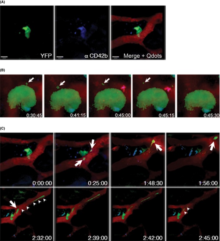
In this review paper, we give a historical perspective of the development of imaging modalities to visualize platelet biogenesis and how this contributed to our current understanding of megakaryopoiesis and thrombopoiesis. We provide some insight how distinct in vivo and in situ imaging methods, including ultramicrographs, have contributed to the current concepts of platelet formation. The onset of intravital microscopy into the mouse bone marrow has markedly modified and challenged our thinking of platelet biogenesis during the last decade. Finally, we discuss ongoing work, which was presented at the recent International Society on Thrombosis and Haemostasis (ISTH) meeting.
Keywords: bone marrow; imaging; megakaryocyte; platelet biogenesis; proplatelets.
Figures
References
-
- Howell WH. Observations upon the occurrence, structure, and function of the giant cells of the marrow. J Morphol. 1890;4:117–130.
-
- Wright J. The origin and nature of blood platelets. Boston Med Surg J. 1906;154:643–645.
-
- Duke WW. The relation of blood platelets to hemorrhagic disease. JAMA. 1910;55:1185–1192. - PubMed
-
- Kuter DJ, Rosenberg RD. The reciprocal relationship of thrombopoietin (c‐Mpl ligand) to changes in the platelet mass during busulfan‐induced thrombocytopenia in the rabbit. Blood. 1995;85:2720–2730. - PubMed
Publication types
LinkOut - more resources
Full Text Sources
Other Literature Sources
