Review
. 2019 Apr;25(2):113-125.
doi: 10.1177/1073858418791763. Epub 2018 Jul 26.

New Insights into the Neurobiology of Restless Legs Syndrome

Affiliations
Review

New Insights into the Neurobiology of Restless Legs Syndrome

Sergi Ferré et al. Neuroscientist. 2019 Apr.

Abstract

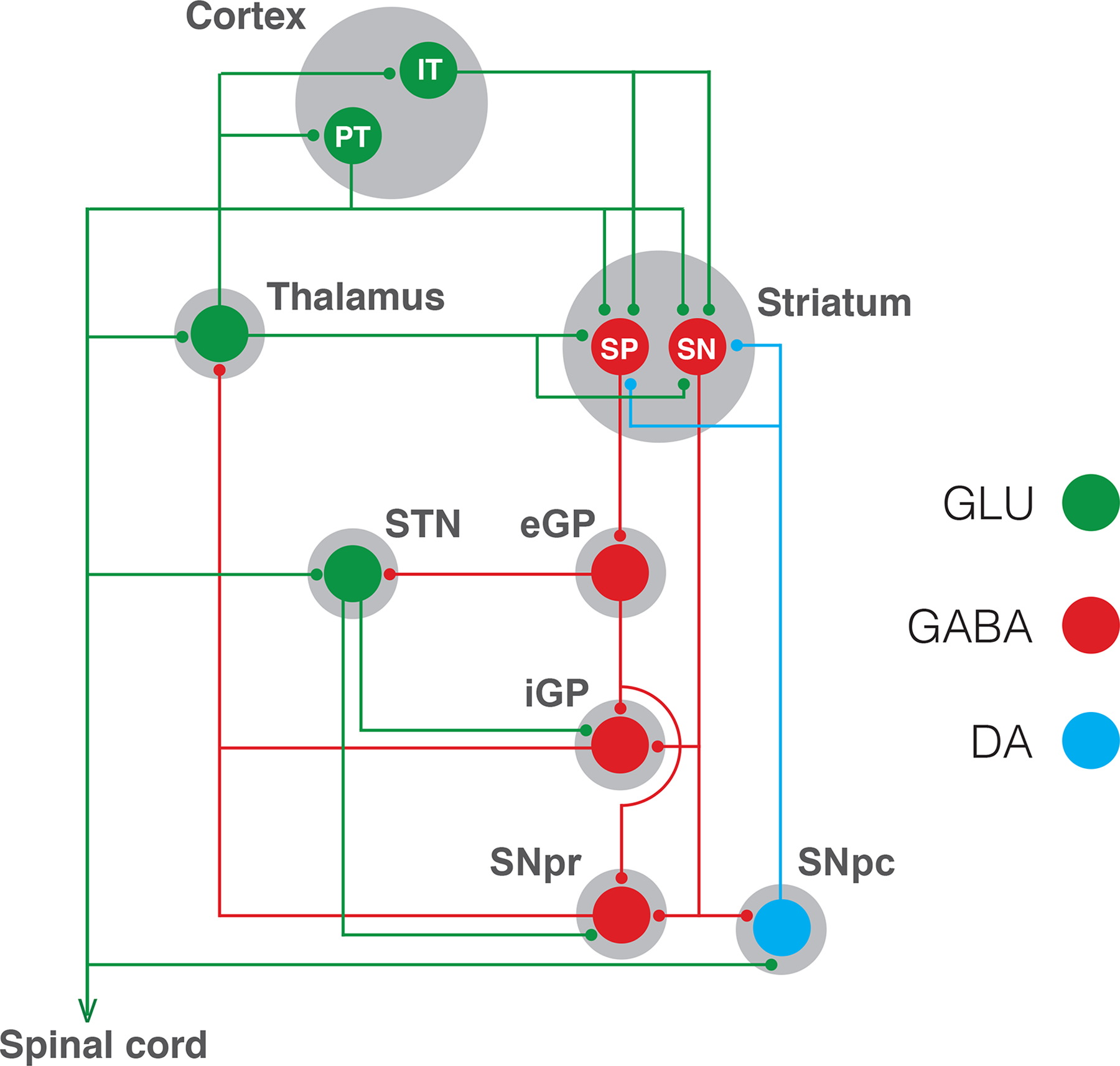
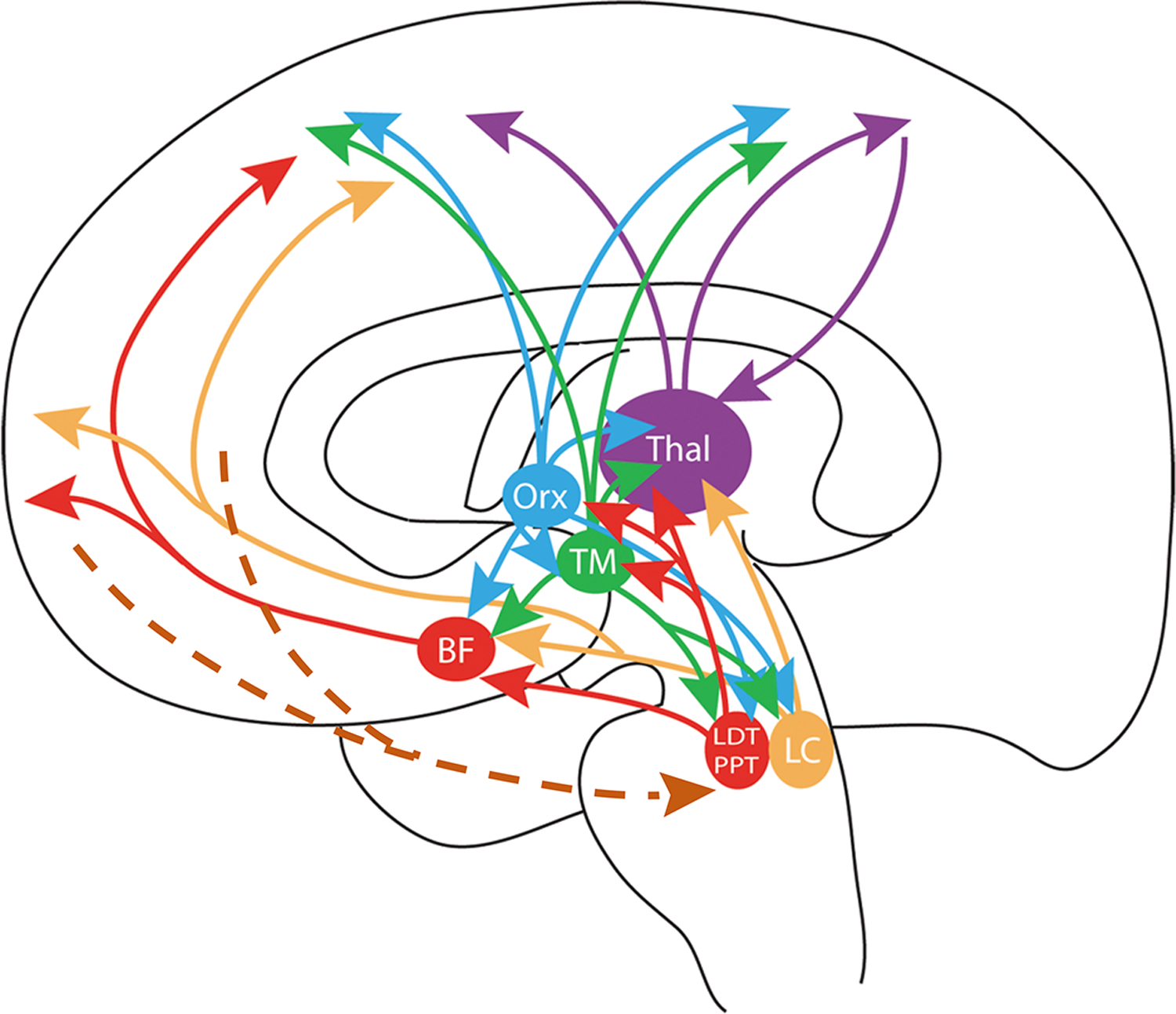
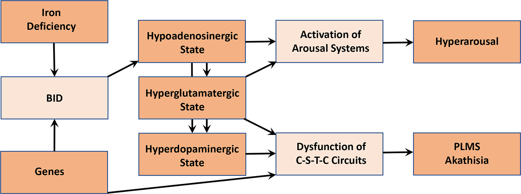
Restless legs syndrome (RLS) is a common sensorimotor disorder, whose basic components include a sensory experience, akathisia, and a sleep-related motor sign, periodic leg movements during sleep (PLMS), both associated with an enhancement of the individual's arousal state. The present review attempts to integrate the major clinical and experimental neurobiological findings into a heuristic pathogenetic model. The model also integrates the recent findings on RLS genetics indicating that RLS has aspects of a genetically moderated neurodevelopmental disorder involving mainly the cortico-striatal-thalamic-cortical circuits. Brain iron deficiency (BID) remains the key initial pathobiological factor and relates to alterations of iron acquisition by the brain, also moderated by genetic factors. Experimental evidence indicates that BID leads to a hyperdopaminergic and hyperglutamatergic states that determine the dysfunction of cortico-striatal-thalamic-cortical circuits in genetically vulnerable individuals. However, the enhanced arousal mechanisms critical to RLS are better explained by functional changes of the ascending arousal systems. Recent experimental and clinical studies suggest that a BID-induced hypoadenosinergic state provides the link for a putative unified pathophysiological mechanism for sensorimotor signs of RLS and the enhanced arousal state.

Keywords: adenosine; arousal; brain iron deficiency; dopamine; glutamate; restless legs syndrome.


Conflict of interest statement

Declaration of Conflicting Interests

The authors declare no conflict of interests

Figures

Figure 1.
Figure 1.
Parallel cortico-striatal-thalamic-cortical circuitry. Two types of pyramidal neurons project from the cortex to the striatum: the intratelencephalic (IT) neuron, which project ipsilaterally and bilaterally to the cortex and striatum; and the pyramidal tract (PT) neuron, which projects ipsilaterally to the striatum, but also projects to the ipsilateral thalamus, subthalamic nucleus (STN), mesencephalon (including the substantia nigra pars compacta, SNpc) and spinal cord. IT and PT neurons project to both subtypes of GABAergic striatal efferent neurons: the striatonigral (SN) neuron, which directly connect the striatum with the output structures of the basal ganglia, which are the substantia nigra pars reticulata (SNpr) and the internal segment of the globus pallidus (iGP); and the striatopallidal (SP) neuron, which connect indirectly with the output structures by a relay through the external segment of the globus pallidus (eGP) and the STN. This scheme shows only the characteristic parallel processing of the basal ganglia, but there is also a substantial level of convergent integration of cortico-striatal projections, more specifically from reciprocally connected cortical regions, such as the motor and somatosensory cortical areas. Dysfunction of cortico-striatal-thalamic-cortical circuitry involving motor and somatosensory cortical areas seem to determine the PLMS and akathia components of RLS.
Figure 2.
Figure 2.
Reduction of the enhancer activity of an RLS-risk common polymorphism of the regulatory locus of the gene MEIS1 in the mouse ganglionic eminence. A. Transgenic mouse embryos after injection of the protective (left, ‘A’ for alanine) and risk allele constructs (right, ‘G’ for guanine). The blue color indicates the regions expressing the reporter gene beta-galactosidase, and the arrowheads indicate the telencephalic signal in the ganglionic eminence. B. Analysis of the signal intensity and volume demonstrated a significant reduction (*) of the enhancer activity of the risk versus the protective construct, down to 35% and 24%, respectively. Reproduced and modified with permission from Spieler and others 2014.
Figure 3.
Figure 3.
Iron deficiency in choroid plexus tissue from RLS patients. Upper, middle and lower micrographs represent iron, heavy-chain ferritin (H-ferritin) and transferrin receptor staining, respectively, from RLS (left panels) and control subjects (middle panels). Right panels represent the respective quantitative analysis, showing a very significant decrease of iron and H-ferritin densities (***) and a significant increase in transferrin receptor density (***) in RLS (left scatterplots) versus control subjects (right scatterplots). Reproduced and modified from Connor and others 2011.
Figure 4.
Figure 4.
Schematic representation of a cortico-striatal glutamatergic terminal and its modulatory dopamine and adenosine receptors. Dopamine and adenosine modulate cortico-striatal glutamate (GLU) release by acting on A1R-A2AR and D2R-D4R heteromers. The A1R-A2AR heteromer acts as an adenosine concentration-dependent switch, by which a low adenosine concentration activates preferentially A1R, which produces inhibition of glutamate release, and a high adenosine concentration also activates A2AR, which shuts down A1R signaling and promotes and A2AR-mediated stimulation of glutamate release. The D2R-D4R heteromer provides a dopamine concentration-dependent stepwise inhibitory mechanism of glutamate release, that depends on the higher affinity of dopamine for the D4R and on a D4R-mediated-increase of D2R signaling. The BID-dependent increase in the excitability of the glutamatergic terminal to release glutamate seems to depend, predominantly, on functional downregulation of A1R, which can be counteracted by D2R or D4R agonists and α2δ ligands (see text). The function of voltage-dependent calcium channels (VDCC), which activation promotes vesicular fusion and neurotransmitter release, is regulated by Gi-coupled receptors, including A1R, D2R and D4R, as well as by accessory α2δ subunits, the targets of gabapentin-like compounds. Reproduced and modified from Yepes and others 2017.
Figure 5.
Figure 5.
Scheme of the highly interconnected multiple ascending arousal systems, which are directly or indirectly involved in the homeostatic sleep function of adenosine. This homeostatic function depends mostly on the accumulation of extracellular adenosine in basal forebrain and cortex. BF: area of origin of the corticopetal basal forebrain system; LC: locus coeruleus; LDT and PPT: laterodorsal and pedunculopontine nuclei; TM: tuberomammillary nucleus; Orx: area of origin of the hypocretin/orexin system; Thal: thalamus. The brown broken lines indicate the existence of descending prefrontal corticofugal neurons that influence the pontomesencephalic tegmentum (see text). Reproduced and modified from Ferré 2010
Figure 6.
Figure 6.
Integrative scheme of the pathogenetic mechanisms involved in the PLMS, akathisia and arousal components of RLS (see text: Conclusion and future directions).

References

    1. Aksu M, Bara-Jimenez W. 2002. State dependent excitability changes of spinal flexor reflex in patients with restless legs syndrome secondary to chronic renal failure. Sleep Med 3:427–30. - PubMed
    1. Allen RP, Mignot E, Ripley B, Nishino S, Earley CJ. 2002. Increased CSF hypocretin-1 (orexin-A) in restless legs syndrome. Neurology 59:639–41. - PubMed
    1. Allen RP, Walters AS, Montplaisir J, Hening W, Myers A, Bell TJ, and others. 2005. Restless legs syndrome prevalence and impact: REST general population study. Arch Intern Med 165:1286–92. - PubMed
    1. Allen RP, Earley CJ. 2007. The role of iron in restless legs syndrome. Mov Disord 18:S440–8. - PubMed
    1. Allen RP, Connor JR, Hyland K, Earley CJ. 2009. Abnormally increased CSF 3-Ortho-methyldopa (3-OMD) in untreated restless legs syndrome (RLS) patients indicates more severe disease and possibly abnormally increased dopamine synthesis. Sleep Med 10:123–8. - PMC - PubMed

Publication types