. 2018 Oct;268(4):564-573.
doi: 10.1097/SLA.0000000000002977.

Xenoantigen Deletion and Chemical Immunosuppression Can Prolong Renal Xenograft Survival

Affiliations

Xenoantigen Deletion and Chemical Immunosuppression Can Prolong Renal Xenograft Survival

Andrew B Adams et al. Ann Surg. 2018 Oct.

Abstract

Objective: Xenotransplantation using pig organs could end the donor organ shortage for transplantation, but humans have xenoreactive antibodies that cause early graft rejection. Genome editing can eliminate xenoantigens in donor pigs to minimize the impact of these xenoantibodies. Here we determine whether an improved cross-match and chemical immunosuppression could result in prolonged kidney xenograft survival in a pig-to-rhesus preclinical model.

Methods: Double xenoantigen (Gal and Sda) knockout (DKO) pigs were created using CRISPR/Cas. Serum from rhesus monkeys (n = 43) was cross-matched with cells from the DKO pigs. Kidneys from the DKO pigs were transplanted into rhesus monkeys (n = 6) that had the least reactive cross-matches. The rhesus recipients were immunosuppressed with anti-CD4 and anti-CD8 T-cell depletion, anti-CD154, mycophenolic acid, and steroids.

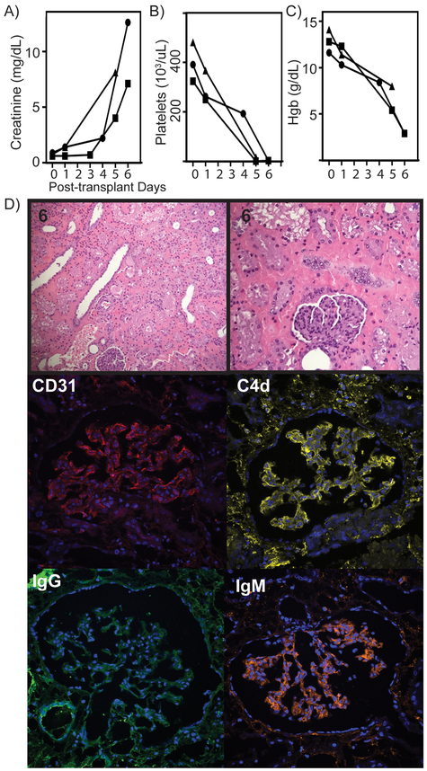
Results: Rhesus antibody binding to DKO cells is reduced, but all still have positive CDC and flow cross-match. Three grafts were rejected early at 5, 6, and 6 days. Longer survival was achieved in recipients with survival to 35, 100, and 435 days. Each of the 3 early graft losses was secondary to IgM antibody-mediated rejection. The 435-day graft loss occurred secondary to IgG antibody-mediated rejection.

Conclusions: Reducing xenoantigens in donor pigs and chemical immunosuppression can be used to achieve prolonged renal xenograft survival in a preclinical model, suggesting that if a negative cross-match can be obtained for humans then prolonged survival could be achieved.


Figures

FIGURE 1.
FIGURE 1.
Genotype and phenotype of double KO pig. (A) GGTA1 and (B) B4GalNT2 gene PCR product noted in wild type, lane 3 (arrow), but absent following Cas9 disruption, lanes 1 and 2. Top band shows internal control, actin PCR product, along top and alone in lane 4. (C) To demonstrate absence of targeted gene activity, flow cytometric PBMC phenotype of double KO pig is stained with IB4 lectin (binding αGal) and DBA lectin (binding SDa antigen). The bottom pair of histograms demonstration of overlay with cells alone controls. (D) Confocal microscopy of renal tissue from wild type and double KO tissue. Top row in wild type and KO pigs respectively, show IB4 and DBA stains alone and bottom row shows overlay with dapi nuclear stain. Images capture a glomerulus. White bar indicates 50uM.
FIGURE 2.
FIGURE 2.
Rhesus Macaque IgG and IgM antibody binding to pig PBMC. (A) Flow cytometric histograms showing IgG, top row, and IgM, bottom row, binding to three genetically modified pig backgrounds. Each representative histogram is overlaid on secondary alone in gray. (B) Flow cytometric MFI of IgG and IgM antibody binding of a group of normal Rhesus Macaque animals (n=14) is shown along X axis and Y axis respectively. GGTA1, GGTA1/B4GalNT2, and lastly GGTA1/CMAH/B4GalNT PBMC antibody binding is demonstrated. Box in left lower corner demonstrates region of estimated negative crossmatch.
FIGURE 3.
FIGURE 3.
Flow cytometric crossmatch and immunosuppression protocol employed in transplants. (A) IgG and IgM flow cytometric MFI and demonstrated for six recipients compared to a subset of screened Rhesus Macaques. All recipients were selected for low pre-transplant IgG antibody binding, but none achieved negative crossmatch as demonstrated by black dotted line. (B) Immunosuppression regimen used in all of the recipients.
FIGURE 4.
FIGURE 4.
Laboratory and tissue analysis of kidney recipients with graft failure in less than one week. (A) Creatinine shown for three recipients of this group showing elevation and graft failure by day 6. (B) Significant thrombocytopenia was developed in all recipients as shown by platelet count. (C) Severe anemia developed quickly in this subset of recipients as shown by hemoglobin levels during first week. (D) H&E stain at low and high magnification of representative tissue. Confocal microscopy of representative glomeruli showing, CD31 endothelial marker, C4d complement activation product, IgG and IgM binding. Significant IgM and C4d binding was noted in all grafts.
FIGURE 5.
FIGURE 5.
Laboratory and tissue analysis of grafts surviving 35-100 days. (A) Creatinine is shown to be well controlled until acutely rising at time of graft failure. (B) Following initial challenge, there is minimal significant thrombocytopenia noted during course of the transplants. (C) H&E of 100 day explant graft tissue. Confocal microscopy showing tissue CD31, C4d, IgG and IgM deposition in the (D) 35 day explant and (E.) and 100 day explant.
FIGURE 6.
FIGURE 6.
Life-supporting renal function and histology for graft living 435 days. (A) Creatinine was adequately cleared for over one year following transplant and then trended upward. (B) Following first week thrombocytopenia, normal platelet levels were maintained over course of graft following. (C) Unlike early graft failure recipients, this graft did not encounter anemia and shown by hemoglobin levels. (D) H&E biopsies from day 170, 240 and explanted tissue at day 435 are shown. Confocal microscopy at time of explant shows CD31, C4d, IgG and IgM staining. (E.) Proteinuria was not encountered as shown by urine protein.

References

    1. Patel R, Terasaki PI. Significance of the positive crossmatch test in kidney transplantation. N Engl J Med. 1969;280(14):735–9. - PubMed
    1. Starzl TE, Marchioro TL, Terasaki PI, Porter KA, Faris TD, Herrmann TJ, et al. Chronic survival after human renal homotransplantation. Lymphocyte-antigen matching, pathology and influence of thymectomy. Ann Surg. 1965;162(4):749–87. - PMC - PubMed
    1. Rosenthal JT, Hakala TR, Iwatsuki S, Shaw BW Jr., Starzl TE. Cadaveric renal transplantation under cyclosporine-steroid therapy. Surg Gynecol Obstet. 1983;157(4):309–15. - PMC - PubMed
    1. Wijkstrom M, Iwase H, Paris W, Hara H, Ezzelarab M, Cooper DK. Renal xenotransplantation: experimental progress and clinical prospects. Kidney Int. 2017;91(4):790–6. - PMC - PubMed
    1. Cooper DK, Ekser B, Ramsoondar J, Phelps C, Ayares D. The role of genetically engineered pigs in xenotransplantation research. J Pathol. 2016;238(2):288–99. - PMC - PubMed

Publication types